


Abstract
Objectives: The objective is to provide a precise segmentation technique based on ACRF, which can handle the variations between major and minor vessels and reduce the interference present in the model due to overfitting and can provide a high-quality reconstructed image. Therefore, a robust method with statistical properties needs to be presented to enhance the performance of the model. Moreover, a statistical framework is required to classify images precisely.
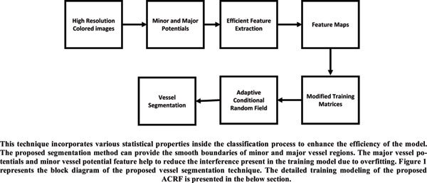
Methods: The Adaptive Conditional Random Field (ACRF) model is used to detect DR disease in the early stages. Here, major vessel potential and minor vessel potential features are extracted for precise segmentation of vessel and non-vessel regions. This feature enhances the efficiency of the model. These major vessel and minor vessel potential features rebuild the retinal vasculature parts precisely and help to capture the contextual information present in the ground truth and label images. This method utilizes an ACRF model to reduce interference and computation complexity. Here, two efficient features are extracted to segment fundus images efficiently, such as major vessel potential and minor vessel potential. The proposed ACRF model can provide the design patterns for both input images and labels with the help of major vessel potential, unlike state-of-arttechniques, which provide patterns for only labels and model the contextual information only in labels, which is essential while performing vessel segmentation.
Results: The performance results are tested on the DRIVE dataset. Experimental results verify the superiority of the proposed vessel segmentation technique based on the ACRF model in terms of accuracy, sensitivity, specificity, and F1-measure and segmentation quality.
Conclusion: A highly efficient vessel segmentation technique is evaluated to describe major and minor vessel regions efficiently based on the ACRF to recognize DR in early stages and to ensure an effective diagnosis using eye Fundus Images. The segmentation process decomposes input images into RGB components through histogram labels based on the proposed ACRF model. Here, the Gabor filtering approach is used for pre-processing and predicting parameters. The proposed segmentation method can provide the smooth boundaries of minor and major vessel regions. The proposed ACRF model can provide the design patterns for both input images and labels with the help of major vessel potentials, unlike state-of-art-techniques, which provide patterns for only labels and model the contextual information only in labels.
Keywords: Vessel segmentation, major vessel potentials, minor vessel potentials, adaptive conditional random field.
Graphical Abstract
[http://dx.doi.org/10.1109/ICECA.2017.8203617]
[http://dx.doi.org/10.1109/ACCESS.2017.2786271]
[http://dx.doi.org/10.1109/DICTA.2017.8227413]
[http://dx.doi.org/10.1109/TMI.2017.2759102] [PMID: 28981409]
[http://dx.doi.org/10.1109/WiSPNET.2017.8299917]
[http://dx.doi.org/10.1109/RBME.2010.2084567] [PMID: 22275207]
[http://dx.doi.org/10.2337/diacare.26.9.2653] [PMID: 12941734]
[http://dx.doi.org/10.2337/diacare.23.3.390] [PMID: 10868871]
[http://dx.doi.org/10.1109/ISBI.2018.8363713]
[http://dx.doi.org/10.1109/IJCNN.2017.7966169]
[http://dx.doi.org/10.1109/MLSP.2016.7738877]
[http://dx.doi.org/10.1109/TBME.2017.2752701] [PMID: 28922110]
[http://dx.doi.org/10.1109/TMI.2004.825627] [PMID: 15084075]
[http://dx.doi.org/10.1109/RTEICT42901.2018.9012438]
Call for Papers in Thematic Issues
?The New Era of Computational Intelligence: Big Data Applications in Health Care?
Analyzing healthcare data has remained a tedious task for data analysts in the current age of research due the nonlinear nature of data. With data sources multiplying and their complexity rising, the most common challenge for medical analysts today is obtaining relevant data for those that need it. The challenge ...read more
Advanced Applications of Artificial Intelligence in Manufacturing Technologies
As one of the most advanced fields of study and technology in existence today, artificial intelligence (AI) is finding more and more applications in production and daily life, especially in the industrial sector. This showcases the many applications of AI in mechanical production, including but not limited to: improving worker ...read more
Advancements in AI and Machine Learning for Enhanced Computer Vision Applications
This special thematic issue is meticulously crafted to provide an extensive exploration of the intricate interplay between Artificial Intelligence (AI) and Machine Learning (ML) within the expansive domain of Computer Vision. The scope of this issue is ambitiously designed to cover a diverse range of topics, incorporating cutting-edge research, advanced ...read more
Advancing Computer Vision and Multimedia Communication for Seamless Human-Machine Interaction
The rapid advancements in computer vision and multimedia communication technologies are revolutionizing the way humans interact with machines. These technologies have the potential to enable seamless and natural human-machine interaction, creating new possibilities for communication, collaboration, and entertainment. The findings will have a significant impact on the development of new ...read more


Related Books

IoT-enabled Sensor Networks: Architecture, Methodologies, Security, and Futuristic Applications

AIoT and Big Data Analytics for Smart Healthcare Applications

Research Trends in Artificial Intelligence: Internet of Things

Artificial Intelligence and Multimedia Data Engineering

Basics of Python Programming: A Quick Guide for Beginners

Blockchain Technology in Healthcare - Concepts, Methodologies, and Applications

Artificial Intelligence and Knowledge Processing: Methods and Applications
Handbook of Artificial Intelligence

Amazon Web Services: the Definitive Guide for Beginners and Advanced Users

The Role of AI in Enhancing IoT-Cloud Applications
 19
19 2
2