Generic placeholder image

Current Signal Transduction Therapy

Editor-in-Chief

ISSN (Print): 1574-3624
ISSN (Online): 2212-389X

Research Article

Evaluation of Fetal Head Circumference (HC) and Biparietal Diameter (BPD (Biparietal Diameter)) in Ultrasound Images Using Multi-Task Deep Convolutional Neural Network

Author(s): Fathimuthu Joharah and Kother Mohideen*

Volume 17, Issue 3, 2022

Published on: 20 August, 2022

Article ID: e130522204772 Pages: 10

DOI: 10.2174/1574362417666220513151926

Price: $65

Abstract

Introduction: Ultrasound imaging is a standard examination during pregnancy that can measure specific biometric parameters towards prenatal diagnosis and estimating gestational age. Fetal head circumference (HC) is a significant factor in determining fetus growth and health.

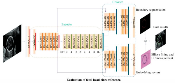
Methods: This paper proposes a multi-task deep convolutional neural network for automatic segmentation and estimation of HC (Fetal head circumference) ellipse by minimizing a compound cost function composed of segmentation dice score and MSE of ellipse parameters. Ultrasoundbased fetal biometric measurements, such as head circumference (HC) and biparietal diameter (BPD (Biparietal Diameter)), are commonly used to evaluate the gestational age and diagnose fetal central nervous system (CNS) pathology. Since manual measurements are operator-dependent and time-consuming, there have been numerous researches on automated methods. However, existing computerized methods still are not satisfactory in terms of accuracy and reliability, owing to difficulties in dealing with various artefacts in ultrasound images.

Results: This paper focuses on fetal head biometry and develops a deep-learning-based method for estimating HC (Fetal head circumference) and BPD (Biparietal Diameter) with a high degree of accuracy and reliability.

Conclusion: The proposed method effectively identifies the head boundary by differentiating tissue image patterns concerning the ultrasound propagation direction. The proposed method was trained with 102 labelled data set and tested to 70 ultrasound images. We achieved a success rate of 92.31% for HC (Fetal head circumference) and BPD (Biparietal Diameter) estimations and an accuracy of 87.14% for the plane acceptance check.

Keywords: Ultrasound images, CNN, MSE, head circumference, biparietal diameter, fetal biometry.

Graphical Abstract
[1]
Abuhamad A, Falkensammer P, Reichartseder F, Zhao Y. Automated retrieval of standard diagnostic fetal cardiac ultrasound planes in the second trimester of pregnancy: A prospective evaluation of software. Ultrasound Obstet Gynecol 2008; 31(1): 30-6.
[http://dx.doi.org/10.1002/uog.5228] [PMID: 18098347]
[2]
Baumgartner CF, Kamnitsas K, Matthew J, Smith S, Kainz B, Rueckert D. Real-time standard scan plane detection and localisation in fetal ultrasound using fully convolutional neural networks. International Conference on Medical Image Computing and Computer-Assisted Intervention, MICCAI: Medical Image Computing and Computer-Assisted Intervention-MICCAI. 2016; pp. 203-11.
[http://dx.doi.org/10.1007/978-3-319-46723-8_24]
[3]
Boykov Y, Veksler O, Zabih R. Fast approximate energy minimization via graph cuts. IEEE Trans Pattern Anal Mach Intell 2001; 23(11): 1222-39.
[http://dx.doi.org/10.1109/34.969114]
[4]
Espinoza J, Good S, Russell E, Lee W. Does the use of automated fetal biometry improve clinical work flow efficiency? J Ultrasound Med 2013; 32(5): 847-50.
[http://dx.doi.org/10.7863/jum.2013.32.5.847] [PMID: 23620327]
[5]
Carneiro G, Georgescu B, Good S, Comaniciu D. Detection and measurement of fetal anatomies from ultrasound images using a con-strained probabilistic boosting tree. IEEE Trans Med Imaging 2008; 27(9): 1342-55.
[http://dx.doi.org/10.1109/TMI.2008.928917] [PMID: 18753047]
[6]
Li J, Wang Y, Lei B, et al. Automatic fetal head circumference measurement in ultrasound using random forest and fast ellipse fitting. IEEE J Biomed Health Inform 2018; 22(1): 215-23.
[http://dx.doi.org/10.1109/JBHI.2017.2703890] [PMID: 28504954]
[7]
Pathak SD, Chalana V, Haynor DR, Kim Y. Edge-guided boundary delineation in prostate ultrasound images. IEEE Trans Med Imaging 2000; 19(12): 1211-9.
[http://dx.doi.org/10.1109/42.897813] [PMID: 11212369]
[8]
Rueda S, Fathima S, Knight CL, et al. Evaluation and comparison of current fetal ultrasound image segmentation methods for biometric measurements: A grand challenge. IEEE Trans Med Imaging 2014; 33(4): 797-813.
[http://dx.doi.org/10.1109/TMI.2013.2276943] [PMID: 23934664]
[9]
Yu J, Wang Y, Chen P. Fetal ultrasound image segmentation system and its use in fetal weight estimation. Med Biol Eng Comput 2008; 46(12): 1227-37.
[http://dx.doi.org/10.1007/s11517-008-0407-y] [PMID: 18850125]
[10]
Hadlock FP, Deter RL, Harrist RB, Park SK. Fetal biparietal diameter: A critical re-evaluation of the relation to menstrual age by means of real-time ultrasound. J Ultrasound Med 1982; 1(3): 97-104.
[http://dx.doi.org/10.7863/jum.1982.1.3.97] [PMID: 6152941]
[11]
Wu L, Cheng J-Z, Li S, Lei B, Wang T, Ni D. FUIQA: Fetal ultrasound image quality assessment with deep convolutional networks. IEEE Trans Cybern 2017; 47(5): 1336-49.
[http://dx.doi.org/10.1109/TCYB.2017.2671898] [PMID: 28362600]
[12]
Ponomarev GV, Gelfand MS, Kazanov MD. A multilevel thresholding combined with edge detection and shape-based recognition for segmentation of fetal ultrasound images. Proc Chall US biometric Meas from fetal ultrasound images (ISBI) 2012; 2012: 17-9.
[13]
Zeng Y, Tsui P-H, Wu W, Zhou Z, Wu S. Fetal ultrasound image segmentation for automatic head circumference biometry using deeply supervised attention-gated V-net. J Digit Imaging 2021; 34(1): 134-48.
[http://dx.doi.org/10.1007/s10278-020-00410-5] [PMID: 33483862]
[14]
Stebbing RV, McManigle JE. A boundary fragment model for head segmentation in fetal ultrasound. Proc Chall US Biometric Meas from Fetal Ultrasound Images ISBI 2012; 9-11.
[15]
Foi A, Maggioni M, Pepe A, et al. Difference of Gaussians revolved along elliptical paths for ultrasound fetal head segmentation. Comput Med Imaging Graph 2014; 38(8): 774-84.
[http://dx.doi.org/10.1016/j.compmedimag.2014.09.006] [PMID: 25450760]
[16]
Shepard M, Filly RA. A standardized plane for biparietal diameter measurement. J Ultrasound Med 1982; 1(4): 145-50.
[http://dx.doi.org/10.7863/jum.1982.1.4.145] [PMID: 6152944]
[17]
Nelson P, Nugent R. The association between sonographic fetal head circumference, obstetric anal sphincter injury and mode of delivery: A retrospective cohort study. Aust New Zeal J Obstet Gynaecol 2021; 61(5): 722-7.
[http://dx.doi.org/10.1111/ajo.13342]
[18]
Gabbay-Benziv R, Anabusi S, Nadir E, Kamar D, Hallak M, Shrim A. The accuracy of sonographic fetal head circumference in twin preg-nancies. J Obstet Gynaecol Can 2021; 43(10): 1159-63.
[http://dx.doi.org/10.1016/j.jogc.2021.02.114] [PMID: 33621678]
[19]
Uddin MS, Halder KK, Tahtali M, et al. Intelligent estimation of noise and blur variances using ANN for the restoration of ultrasound images. Appl Opt 2016; 55(31): 8905-15.
[http://dx.doi.org/10.1364/AO.55.008905] [PMID: 27828292]
[20]
Zhang J, Petitjean C, Yger F, Ainouz S. Explainability for regression CNN in fetal head circumference estimation from ultrasound images. In: Interpretable and Annotation-Efficient Learning for Medical Image Computing. Springer 2020; pp. 73-82.
[http://dx.doi.org/10.1007/978-3-030-61166-8_8]
[21]
Kessler J, Johnsen SL, Ebbing C, Karlsen HO, Rasmussen S, Kiserud T. Estimated date of delivery based on second trimester fetal head circumference: A population-based validation of 21 451 deliveries. Acta Obstet Gynecol Scand 2019; 98(1): 101-5.
[http://dx.doi.org/10.1111/aogs.13454] [PMID: 30168856]
[22]
Loughna P, Chitty L, Evans T, Chudleigh T. Fetal size and dating: Charts recommended for clinical obstetric practice. Ultrasound 2009; 17(3): 160-6.
[http://dx.doi.org/10.1179/174313409X448543]
[23]
Elbah M, Yallese MA, Aouici H, Mabrouki T, Rigal J-F. Comparative assessment of wiper and conventional ceramic tools on surface roughness in hard turning AISI 4140 steel. Meas 2013; 46(9): 3041-56.
[http://dx.doi.org/10.1016/j.measurement.2013.06.018]
[24]
Ciurte A, Bresson X, Cuadra MB. A semi-supervised patch-based approach for segmentation of fetal ultrasound imaging. Proc. Chall. US Biometric Meas from Fetal Ultrasound Images 2012; 2012: 5-7.
[25]
Lu W, Tan J, Floyd R. Automated fetal head detection and measurement in ultrasound images by iterative randomized Hough transform. Ultrasound Med Biol 2005; 31(7): 929-36.
[http://dx.doi.org/10.1016/j.ultrasmedbio.2005.04.002] [PMID: 15972198]
[26]
WisnuJatmiko IPS. Habibie I, Anwar Ma’sum M, Rahmatullah R. Automated telehealth system for fetal growth detection and approxima-tion of ultrasound images. Int J Smart Sensing Intell Syst 2015; 8: 697-719.
[27]
Perez-Gonzalez JL, Muńoz JCB, Porras MCR, Arámbula-Cosío F, Medina-Bańuelos V. Automatic fetal head measurements from ultrasound images using optimal ellipse detection and texture maps, VI Latin American Congress on Biomedical Engineering CLAIB 2014. Paraná, Argentina 29, 30 & 31 October 2014 2015; pp. 329-.
[http://dx.doi.org/10.1007/978-3-319-13117-7_85]
[28]
Meyer R, Rottenstreich A, Shapira M, et al. The role of fetal head circumference in the formation of obstetric anal sphincter injuries fol-lowing vacuum deliveries among primiparous women. Arch Gynecol Obstet 2020; 301(6): 1423-9.
[http://dx.doi.org/10.1007/s00404-020-05558-7] [PMID: 32363548]
[29]
van den Heuvel TLA, de Bruijn D, de Korte CL, Ginneken BV. Automated measurement of fetal head circumference using 2D ultrasound images. PLoS One 2018; 13(8): e0200412.
[http://dx.doi.org/10.1371/journal.pone.0200412] [PMID: 30138319]
[30]
Avalokita DT, Rismonita T, Handayani A, Setiawan AW. Automatic fetal head circumference measurement in 2D ultrasound images based on optimized fast ellipse fitting In: 2020 IEEE Region 10 Conference. Tencon 2020; pp. 37-42.
[31]
Budd BKS, Sinclair M, Khanal B, et al. Conduent head circumference measurement from ultrasound with real-time feedback for sonog-raphers. MICCAI 2019; 683-91.
[32]
Poojari VG, Jose A, Pai MV. Sonographic estimation of the fetal head circumference: Accuracy and factors affecting the error. J Obstet Gynecol India 2021.
[http://dx.doi.org/10.1007/s13224-021-01574-y]
[33]
He K, Gkioxari G, Dollár P, Girshick R. Mask R-CNN Proceedings of the IEEE International Conference on Computer Vision (ICCV). 2017; pp. 2980-8.
[34]
Wang Y, Liang W, Shen J, Jia Y, Yu L-F. A deep coarse-to-fine network for head pose estimation from synthetic data. Pattern Recognit 2019; 94: 196-206.
[http://dx.doi.org/10.1016/j.patcog.2019.05.026]
[35]
Dubost F, Bortsova G, Adams H, et al. Hydranet: Data Augmentation for Regression Neural Networks. In: Medical Image Computing and Computer Assisted Intervention – MICCAI 2019. MICCAI 2019. Lecture Notes in Computer Science. Springer, Cham 2019; p. 11767.
[http://dx.doi.org/10.1007/978-3-030-32251-9_48]
[36]
Esmaeili A, Marvasti F. A novel approach to quantized matrix completion using huber loss measure. IEEE Signal Process Lett 2019; 26(2): 337-41.
[http://dx.doi.org/10.1109/LSP.2019.2891134]
[37]
He K, Zhang X, Ren S, Sun J. Deep residual learning for image recognition In: Proceedings of the IEEE conference on computer vision and pattern recognition. 2016; pp. 770-8.
[38]
Jardim SM, Figueiredo MAT. Segmentation of fetal ultrasound images. Ultrasound Med Biol 2005; 31(2): 243-50.
[http://dx.doi.org/10.1016/j.ultrasmedbio.2004.11.003] [PMID: 15708464]
[39]
Kim HP, Lee SM, Kwon J-Y, Park Y, Kim KC, Seo JK. Automatic evaluation of fetal head biometry from ultrasound images using ma-chine learning. Physiol Meas 2019; 40(6): 065009.
[http://dx.doi.org/10.1088/1361-6579/ab21ac] [PMID: 31091515]
[40]
Lathuilière S, Mesejo P, Alameda-Pineda X, Horaud R. A comprehensive analysis of deep regression. IEEE Trans Pattern Anal Mach Intell 2020; 42(9): 2065-81.
[http://dx.doi.org/10.1109/TPAMI.2019.2910523] [PMID: 30990175]

Rights & Permissions Print Cite
© 2024 Bentham Science Publishers | Privacy Policy