Generic placeholder image

Current Neuropharmacology

Editor-in-Chief

ISSN (Print): 1570-159X
ISSN (Online): 1875-6190

General Review Article

Selecting the Best Animal Model of Parkinson’s Disease for Your Research Purpose: Insight from in vivo PET Imaging Studies

Author(s): Caroline Cristiano Real, Karina Henrique Binda, Majken Borup Thomsen, Thea Pinholt Lillethorup, David James Brooks and Anne Marlene Landau*

Volume 21, Issue 5, 2023

Published on: 16 February, 2023

Page: [1241 - 1272] Pages: 32

DOI: 10.2174/1570159X21666230216101659

Price: $65

Abstract

Parkinson’s disease (PD) is a debilitating neurodegenerative multisystem disorder leading to motor and non-motor symptoms in millions of individuals. Despite intense research, there is still no cure, and early disease biomarkers are lacking. Animal models of PD have been inspired by basic elements of its pathogenesis, such as dopamine dysfunction, alpha-synuclein accumulation, neuroinflammation and disruption of protein degradation, and these have been crucial for a deeper understanding of the mechanisms of pathology, the identification of biomarkers, and evaluation of novel therapies. Imaging biomarkers are non-invasive tools to assess disease progression and response to therapies; their discovery and validation have been an active field of translational research. Here, we highlight different considerations of animal models of PD that can be applied to future research, in terms of their suitability to answer different research questions. We provide the reader with important considerations of the best choice of model to use based on the disease features of each model, including issues related to different species. In addition, positron emission tomography studies conducted in PD animal models in the last 5 years are presented. With a variety of different species, interventions and genetic information, the choice of the most appropriate model to answer research questions can be daunting, especially since no single model recapitulates all aspects of this complex disorder. Appropriate animal models in conjunction with in vivo molecular imaging tools, if selected properly, can be a powerful combination for the assessment of novel therapies and developing tools for early diagnosis.

Keywords: Animal models, Parkinson’s disease, rodent, non-human primate, minipig, alpha-synuclein, positron emission tomography, autoradiography.
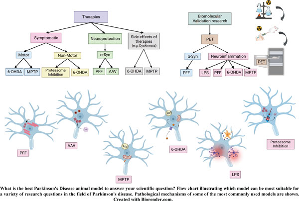
Graphical Abstract
[1]
Poewe, W.; Seppi, K.; Tanner, C.M.; Halliday, G.M.; Brundin, P.; Volkmann, J.; Schrag, A.E.; Lang, A.E. Parkinson disease. Nat. Rev. Dis. Primers, 2017, 3(1), 17013.
[http://dx.doi.org/10.1038/nrdp.2017.13] [PMID: 28332488]
[2]
Postuma, R.B.; Berg, D.; Stern, M.; Poewe, W.; Olanow, C.W.; Oertel, W.; Obeso, J.; Marek, K.; Litvan, I.; Lang, A.E.; Halliday, G.; Goetz, C.G.; Gasser, T.; Dubois, B.; Chan, P.; Bloem, B.R.; Adler, C.H.; Deuschl, G. MDS clinical diagnostic criteria for Parkinson’s disease. Mov. Disord., 2015, 30(12), 1591-1601.
[http://dx.doi.org/10.1002/mds.26424] [PMID: 26474316]
[3]
Hirsch, E.C.; Standaert, D.G. Ten unsolved questions about neuroinflammation in Parkinson’s disease. Mov. Disord., 2021, 36(1), 16-24.
[http://dx.doi.org/10.1002/mds.28075] [PMID: 32357266]
[4]
Scheperjans, F.; Aho, V.; Pereira, P.A.B.; Koskinen, K.; Paulin, L.; Pekkonen, E.; Haapaniemi, E.; Kaakkola, S.; Eerola-Rautio, J.; Pohja, M.; Kinnunen, E.; Murros, K.; Auvinen, P. Gut microbiota are related to Parkinson’s disease and clinical phenotype. Mov. Disord., 2015, 30(3), 350-358.
[http://dx.doi.org/10.1002/mds.26069] [PMID: 25476529]
[5]
Jo, S.; Kim, Y.J.; Park, K.W.; Hwang, Y.S.; Lee, S.H.; Kim, B.J.; Chung, S.J. Association of NO2 and other air pollution exposures with the risk of parkinson disease. JAMA Neurol., 2021, 78(7), 800-808.
[http://dx.doi.org/10.1001/jamaneurol.2021.1335] [PMID: 33999109]
[6]
Yan, D.; Zhang, Y.; Liu, L.; Shi, N.; Yan, H. Pesticide exposure and risk of Parkinson’s disease: Dose-response meta-analysis of observational studies. Regul. Toxicol. Pharmacol., 2018, 96, 57-63.
[http://dx.doi.org/10.1016/j.yrtph.2018.05.005] [PMID: 29729297]
[7]
Smith, L.; Schapira, A.H.V. GBA variants and Parkinson disease: Mechanisms and treatments. Cells, 2022, 11(8), 1261.
[http://dx.doi.org/10.3390/cells11081261] [PMID: 35455941]
[8]
Schrag, A.; Horsfall, L.; Walters, K.; Noyce, A.; Petersen, I. Prediagnostic presentations of Parkinson’s disease in primary care: A case-control study. Lancet Neurol., 2015, 14(1), 57-64.
[http://dx.doi.org/10.1016/S1474-4422(14)70287-X] [PMID: 25435387]
[9]
Greenland, J.C.; Williams-Gray, C.H.; Barker, R.A. The clinical heterogeneity of Parkinson’s disease and its therapeutic implications. Eur. J. Neurosci., 2019, 49(3), 328-338.
[http://dx.doi.org/10.1111/ejn.14094] [PMID: 30059179]
[10]
Berg, D.; Borghammer, P.; Fereshtehnejad, S.M.; Heinzel, S.; Horsager, J.; Schaeffer, E.; Postuma, R.B. Prodromal Parkinson disease subtypes — key to understanding heterogeneity. Nat. Rev. Neurol., 2021, 17(6), 349-361.
[http://dx.doi.org/10.1038/s41582-021-00486-9] [PMID: 33879872]
[11]
Borghammer, P.; Horsager, J.; Andersen, K.; Van Den Berge, N.; Raunio, A.; Murayama, S.; Parkkinen, L.; Myllykangas, L. Neuropathological evidence of body-first vs. brain-first Lewy body disease. Neurobiol. Dis., 2021, 161, 105557.
[http://dx.doi.org/10.1016/j.nbd.2021.105557] [PMID: 34763110]
[12]
Horsager, J.; Knudsen, K.; Sommerauer, M. Clinical and imaging evidence of brain-first and body-first Parkinson’s disease. Neurobiol. Dis., 2022, 164, 105626.
[http://dx.doi.org/10.1016/j.nbd.2022.105626] [PMID: 35031485]
[13]
Hutny, M.; Hofman, J.; Klimkowicz-Mrowiec, A.; Gorzkowska, A. Current knowledge on the background, pathophysiology and treatment of levodopa-induced dyskinesia—literature review. J. Clin. Med., 2021, 10(19), 4377.
[http://dx.doi.org/10.3390/jcm10194377] [PMID: 34640395]
[14]
Pahwa, R. Amantadine: An old drug reborn. Lancet Neurol., 2021, 20(12), 975-977.
[http://dx.doi.org/10.1016/S1474-4422(21)00356-2] [PMID: 34678172]
[15]
Hacker, M.; Cannard, G.; Turchan, M.; Meystedt, J.; Davis, T.; Phibbs, F.; Hedera, P.; Konrad, P.; Charles, D. Early subthalamic nucleus deep brain stimulation in Parkinson’s disease reduces long-term medication costs. Clin. Neurol. Neurosurg., 2021, 210, 106976.
[http://dx.doi.org/10.1016/j.clineuro.2021.106976] [PMID: 34666273]
[16]
Sharma, V.D.; Patel, M.; Miocinovic, S. Surgical treatment of Parkinson’s disease: Devices and lesion approaches. Neurotherapeutics, 2020, 17(4), 1525-1538.
[http://dx.doi.org/10.1007/s13311-020-00939-x] [PMID: 33118132]
[17]
Nag, N.; Jelinek, G.A. More research is needed on lifestyle behaviors that influence progression of Parkinson’s disease. Front. Neurol., 2019, 10, 452.
[http://dx.doi.org/10.3389/fneur.2019.00452] [PMID: 31114542]
[18]
Mischley, L.K.; Lau, R.C.; Bennett, R.D. Role of diet and nutritional supplements in Parkinson’s disease progression. Oxid. Med. Cell. Longev., 2017, 2017, 1-9.
[http://dx.doi.org/10.1155/2017/6405278] [PMID: 29081890]
[19]
Ferreira, A.F.F.; Binda, K.H.; Real, C.C. The effects of treadmill exercise in animal models of Parkinson’s disease: A systematic review. Neurosci. Biobehav. Rev., 2021, 131, 1056-1075.
[http://dx.doi.org/10.1016/j.neubiorev.2021.10.019] [PMID: 34688727]
[20]
Svensson, M.; Lexell, J.; Deierborg, T. Effects of physical exercise on neuroinflammation, neuroplasticity, neurodegeneration, and behavior. Neurorehabil. Neural Repair, 2015, 29(6), 577-589.
[http://dx.doi.org/10.1177/1545968314562108] [PMID: 25527485]
[21]
Alipour Nosrani, E.; Tamtaji, O.R.; Alibolandi, Z.; Sarkar, P.; Ghazanfari, M.; Azami Tameh, A.; Taghizadeh, M.; Banikazemi, Z.; Hadavi, R.; Naderi Taheri, M. Neuroprotective effects of probiotics bacteria on animal model of Parkinson’s disease induced by 6-hydroxydopamine: A behavioral, biochemical, and histological study. J. Immunoassay Immunochem., 2021, 42(2), 106-120.
[http://dx.doi.org/10.1080/15321819.2020.1833917] [PMID: 33078659]
[22]
Binda, K.H.; Lillethorup, T.P.; Real, C.C.; Bærentzen, S.L.; Nielsen, M.N.; Orlowski, D.; Brooks, D.J.; Chacur, M.; Landau, A.M. Exercise protects synaptic density in a rat model of Parkinson’s disease. Exp. Neurol., 2021, 342, 113741.
[http://dx.doi.org/10.1016/j.expneurol.2021.113741] [PMID: 33965411]
[23]
Real, C.C.; Doorduin, J.; Kopschina Feltes, P.; Vállez García, D.; de Paula Faria, D.; Britto, L.R.; de Vries, E.F.J. Evaluation of exercise-induced modulation of glial activation and dopaminergic damage in a rat model of Parkinson’s disease using [ 11 C]PBR28 and [ 18 F]FDOPA PET. J. Cereb. Blood Flow Metab., 2019, 39(6), 989-1004.
[http://dx.doi.org/10.1177/0271678X17750351] [PMID: 29271291]
[24]
Johansson, M.E.; Cameron, I.G.M.; Van der Kolk, N.M.; Vries, N.M.; Klimars, E.; Toni, I.; Bloem, B.R.; Helmich, R.C. Aerobic exercise alters brain function and structure in Parkinson’s disease: A randomized controlled trial. Ann. Neurol., 2022, 91(2), 203-216.
[http://dx.doi.org/10.1002/ana.26291] [PMID: 34951063]
[25]
Johansson, H.; Freidle, M.; Ekman, U.; Schalling, E.; Leavy, B.; Svenningsson, P.; Hagströmer, M.; Franzén, E. Feasibility aspects of exploring exercise-induced neuroplasticity in Parkinson’s disease: A pilot randomized controlled trial. Parkinsons Dis., 2020, 2020, 1-12.
[http://dx.doi.org/10.1155/2020/2410863] [PMID: 32300475]
[26]
Chromiec, P.A.; Urbaś, Z.K.; Jacko, M.; Kaczor, J.J. The proper diet and regular physical activity slow down the development of Parkinson disease. Aging Dis., 2021, 12(7), 1605-1623.
[http://dx.doi.org/10.14336/AD.2021.0123] [PMID: 34631210]
[27]
Helmich, R.C.; Vaillancourt, D.E.; Brooks, D.J. The future of brain imaging in Parkinson’s disease. J. Parkinsons Dis., 2018, 8(s1), S47-S51.
[http://dx.doi.org/10.3233/JPD-181482] [PMID: 30584163]
[28]
Bidesi, N.S.R.; Vang Andersen, I.; Windhorst, A.D.; Shalgunov, V.; Herth, M.M. The role of neuroimaging in Parkinson’s disease. J. Neurochem., 2021, 159(4), 660-689.
[http://dx.doi.org/10.1111/jnc.15516] [PMID: 34532856]
[29]
Bannon, D.; Landau, A.M.; Doudet, D.J. How relevant are imaging findings in animal models of movement disorders to human disease? Curr. Neurol. Neurosci. Rep., 2015, 15(8), 53.
[http://dx.doi.org/10.1007/s11910-015-0571-z] [PMID: 26092313]
[30]
Bryda, E.C. The Mighty Mouse: The impact of rodents on advances in biomedical research. Mol. Med., 2013, 110(3), 207-211.
[PMID: 23829104]
[31]
Collier, T.J.; Kanaan, N.M.; Kordower, J.H. Aging and Parkinson’s disease: Different sides of the same coin? Mov. Disord., 2017, 32(7), 983-990.
[http://dx.doi.org/10.1002/mds.27037] [PMID: 28520211]
[32]
Van Den Berge, N.; Ferreira, N.; Mikkelsen, T.W.; Alstrup, A.K.O.; Tamgüney, G.; Karlsson, P.; Terkelsen, A.J.; Nyengaard, J.R.; Jensen, P.H.; Borghammer, P. Ageing promotes pathological alpha-synuclein propagation and autonomic dysfunction in wild-type rats. Brain, 2021, 144(6), 1853-1868.
[http://dx.doi.org/10.1093/brain/awab061] [PMID: 33880502]
[33]
Collier, T.J.; O’Malley, J.; Rademacher, D.J.; Stancati, J.A.; Sisson, K.A.; Sortwell, C.E.; Paumier, K.L.; Gebremedhin, K.G.; Steece-Collier, K. Interrogating the aged striatum: Robust survival of grafted dopamine neurons in aging rats produces inferior behavioral recovery and evidence of impaired integration. Neurobiol. Dis., 2015, 77, 191-203.
[http://dx.doi.org/10.1016/j.nbd.2015.03.005] [PMID: 25771169]
[34]
Jackson, S.J.; Andrews, N.; Ball, D.; Bellantuono, I.; Gray, J.; Hachoumi, L.; Holmes, A.; Latcham, J.; Petrie, A.; Potter, P.; Rice, A.; Ritchie, A.; Stewart, M.; Strepka, C.; Yeoman, M.; Chapman, K. Does age matter? The impact of rodent age on study outcomes. Lab. Anim., 2017, 51(2), 160-169.
[http://dx.doi.org/10.1177/0023677216653984] [PMID: 27307423]
[35]
Mari, Z.; Mestre, T.A. The disease modification conundrum in Parkinson’s disease: Failures and hopes. Front. Aging Neurosci., 2022, 14, 810860.
[http://dx.doi.org/10.3389/fnagi.2022.810860] [PMID: 35296034]
[36]
Prensa, L.; Parent, A. The nigrostriatal pathway in the rat: A single-axon study of the relationship between dorsal and ventral tier nigral neurons and the striosome/matrix striatal compartments. J. Neurosci., 2001, 21(18), 7247-7260.
[http://dx.doi.org/10.1523/JNEUROSCI.21-18-07247.2001] [PMID: 11549735]
[37]
Blesa, J.; Trigo-Damas, I.; del Rey, N.L.G.; Obeso, J.A. The use of nonhuman primate models to understand processes in Parkinson’s disease. J. Neural Transm. (Vienna), 2018, 125(3), 325-335.
[http://dx.doi.org/10.1007/s00702-017-1715-x] [PMID: 28357564]
[38]
Emborg, M.E. Nonhuman primate models of Parkinson’s disease. ILAR J., 2007, 48(4), 339-355.
[http://dx.doi.org/10.1093/ilar.48.4.339] [PMID: 17712221]
[39]
Capitanio, J.P.; Emborg, M.E. Contributions of non-human primates to neuroscience research. Lancet, 2008, 371(9618), 1126-1135.
[http://dx.doi.org/10.1016/S0140-6736(08)60489-4] [PMID: 18374844]
[40]
Bjarkam, C.R.; Nielsen, M.S.; Glud, A.N.; Rosendal, F.; Mogensen, P.; Bender, D.; Doudet, D.; Møller, A.; Sørensen, J.C. Neuromodulation in a minipig MPTP model of Parkinson disease. Br. J. Neurosurg., 2008, 22(Suppl. 1), S9-S12.
[http://dx.doi.org/10.1080/02688690802448285] [PMID: 19085346]
[41]
Lind, N.M.; Moustgaard, A.; Jelsing, J.; Vajta, G.; Cumming, P.; Hansen, A.K. The use of pigs in neuroscience: Modeling brain disorders. Neurosci. Biobehav. Rev., 2007, 31(5), 728-751.
[http://dx.doi.org/10.1016/j.neubiorev.2007.02.003] [PMID: 17445892]
[42]
Hoffe, B.; Holahan, M.R. The use of pigs as a translational model for studying neurodegenerative diseases. Front. Physiol., 2019, 10, 838.
[http://dx.doi.org/10.3389/fphys.2019.00838] [PMID: 31354509]
[43]
Bjarkam, C.R.; Glud, A.N.; Orlowski, D.; Sørensen, J.C.H.; Palomero-Gallagher, N. The telencephalon of the Göttingen minipig, cytoarchitecture and cortical surface anatomy. Brain Struct. Funct., 2017, 222(5), 2093-2114.
[http://dx.doi.org/10.1007/s00429-016-1327-5] [PMID: 27778106]
[44]
Pabst, R. The pig as a model for immunology research. Cell Tissue Res., 2020, 380(2), 287-304.
[http://dx.doi.org/10.1007/s00441-020-03206-9] [PMID: 32356014]
[45]
Wernersson, R.; Schierup, M.H.; Jørgensen, F.G.; Gorodkin, J.; Panitz, F.; Stærfeldt, H.H.; Christensen, O.F.; Mailund, T.; Hornshøj, H.; Klein, A.; Wang, J.; Liu, B.; Hu, S.; Dong, W.; Li, W.; Wong, G.K.S.; Yu, J.; Wang, J.; Bendixen, C.; Fredholm, M.; Brunak, S.; Yang, H.; Bolund, L. Pigs in sequence space: A 0.66X coverage pig genome survey based on shotgun sequencing. BMC Genomics, 2005, 6(1), 70.
[http://dx.doi.org/10.1186/1471-2164-6-70] [PMID: 15885146]
[46]
Meyer, W.; Kacza, J.; Zschemisch, N.H.; Godynicki, S.; Seeger, J. Observations on the actual structural conditions in the stratum superficiale dermidis of porcine ear skin, with special reference to its use as model for human skin. Ann. Anat., 2007, 189(2), 143-156.
[http://dx.doi.org/10.1016/j.aanat.2006.09.004] [PMID: 17419547]
[47]
Bailey, M.; Haverson, K.; Inman, C.; Harris, C.; Jones, P.; Corfield, G.; Miller, B.; Stokes, C. The development of the mucosal immune system pre- and post-weaning: Balancing regulatory and effector function. Proc. Nutr. Soc., 2005, 64(4), 451-457.
[http://dx.doi.org/10.1079/PNS2005452] [PMID: 16313686]
[48]
Gutierrez, K.; Dicks, N.; Glanzner, W.G.; Agellon, L.B.; Bordignon, V. Efficacy of the porcine species in biomedical research. Front. Genet., 2015, 6, 293.
[http://dx.doi.org/10.3389/fgene.2015.00293] [PMID: 26442109]
[49]
Mikkelsen, M. MØller, A.; Jensen, L.H.; Pedersen, A.; Harajehi, J.B.; Pakkenberg, H. MPTP-induced Parkinsonism in minipigs: A behavioral, biochemical, and histological study. Neurotoxicol. Teratol., 1999, 21(2), 169-175.
[http://dx.doi.org/10.1016/S0892-0362(98)00037-3] [PMID: 10192277]
[50]
Slot, N.M.; Nørgaard, G.A.; Møller, A.; Mogensen, P.; Bender, D.; Christian, S.J.; Doudet, D.; Reidies Bjarkam, C. Continuous MPTP intoxication in the Göttingen minipig results in chronic parkinsonian deficits. Acta Neurobiol. Exp. (Warsz.), 2016, 76(3), 199-211.
[http://dx.doi.org/10.21307/ane-2017-020] [PMID: 27685773]
[51]
Lillethorup, T.P.; Noer, O.; Alstrup, A.K.O.; Real, C.C.; Stokholm, K.; Thomsen, M.B.; Zaer, H.; Orlowski, D.; Mikkelsen, T.W.; Glud, A.N.; Nielsen, E.H.T.; Schacht, A.C.; Winterdahl, M.; Brooks, D.J.; Sørensen, J.C.H.; Landau, A.M. Spontaneous partial recovery of striatal dopaminergic uptake despite nigral cell loss in asymptomatic MPTP-lesioned female minipigs. Neurotoxicology, 2022, 91, 166-176.
[http://dx.doi.org/10.1016/j.neuro.2022.05.006] [PMID: 35569565]
[52]
Lillethorup, T.P.; Glud, A.N.; Landeck, N.; Alstrup, A.K.O.; Jakobsen, S.; Vang, K.; Doudet, D.J.; Brooks, D.J.; Kirik, D.; Hinz, R.; Sørensen, J.C.; Landau, A.M. In vivo quantification of glial activation in minipigs overexpressing human α-synuclein. Synapse, 2018, 72(12), e22060.
[http://dx.doi.org/10.1002/syn.22060] [PMID: 30009467]
[53]
Lillethorup, T.P.; Glud, A.N.; Alstrup, A.K.O.; Mikkelsen, T.W.; Nielsen, E.H.; Zaer, H.; Doudet, D.J.; Brooks, D.J.; Sørensen, J.C.H.; Orlowski, D.; Landau, A.M. Nigrostriatal proteasome inhibition impairs dopamine neurotransmission and motor function in minipigs. Exp. Neurol., 2018, 303, 142-152.
[http://dx.doi.org/10.1016/j.expneurol.2018.02.005] [PMID: 29428213]
[54]
Lillethorup, T.P.; Glud, A.N.; Alstrup, A.K.O.; Noer, O.; Nielsen, E.H.T.; Schacht, A.C.; Landeck, N.; Kirik, D.; Orlowski, D.; Sørensen, J.C.H.; Doudet, D.J.; Landau, A.M. Longitudinal monoaminergic PET imaging of chronic proteasome inhibition in minipigs. Sci. Rep., 2018, 8(1), 15715.
[http://dx.doi.org/10.1038/s41598-018-34084-5] [PMID: 30356172]
[55]
Larsen, K.; Bæk, R.; Sahin, C.; Kjær, L.; Christiansen, G.; Nielsen, J.; Farajzadeh, L.; Otzen, D.E. Molecular characteristics of porcine alpha-synuclein splicing variants. Biochimie, 2021, 180, 121-133.
[http://dx.doi.org/10.1016/j.biochi.2020.10.019] [PMID: 33152422]
[56]
Dall, A.M.; Danielsen, E.H.; Sørensen, J.C.; Andersen, F.; Møller, A.; Zimmer, J.; Gjedde, A.H.; Cumming, P.; Zimmer, J.; Brevig, T.; Dall, A.M.; Meyer, M.; Pedersen, E.B.; Gjedde, A.; Danielsen, E.H.; Cumming, P.; Andersen, F.; Bender, D.; Falborg, L.; Gee, A.; Gillings, N.M.; Hansen, S.B.; Hermansen, F.; Jørgensen, H.A.; Munk, O.; Poulsen, P.H.; Rodell, A.B.; Sakoh, M.; Simonsen, C.Z.; Smith, D.F.; Sørensen, J.C.; Østergård, L.; Moller, A.; Johansen, T.E. Quantitative [18F]fluorodopa/PET and histology of fetal mesencephalic dopaminergic grafts to the striatum of MPTP-poisoned minipigs. Cell Transplant., 2002, 11(8), 733-746.
[http://dx.doi.org/10.3727/000000002783985314] [PMID: 12588105]
[57]
Danielsen, E.H.; Dimming, P.; Andersen, F.; Bender, D.; Brevig, T.; Falborg, L.; Gee, A.; Gillings, N.M.; Hansen, S.B.; Hermansen, F.; Johansen, J.; Johansen, T.E.; Dahl-Jørgensen, A.; Jørgensen, H.A.; Meyer, M.; Munk, O.; Pedersen, E.B.; Poulsen, P.H.; Rodell, A.B.; Sakoh, M.; Simonsen, C.Z.; Smith, D.F.; Sørensen, J.C.; Østergård, L.; Zimmer, J.; Gjedde, A.; Møller, A. The DaNeX study of embryonic mesencephalic, dopaminergic tissue grafted to a minipig model of Parkinson’s disease: Preliminary findings of effect of MPTP poisoning on striatal dopaminergic markers. Cell Transplant., 2000, 9(2), 247-259.
[http://dx.doi.org/10.1177/096368970000900210] [PMID: 10811397]
[58]
Cumming, P.; Danielsen, E.H.; Vafaee, M.; Falborg, L.; Steffensen, E.; Sørensen, J.C.; Gillings, N.; Bender, D.; Marthi, K.; Andersen, F.; Munk, O.; Smith, D.; Møller, A.; Gjedde, A. Normalization of markers for dopamine innervation in striatum of MPTP-lesioned miniature pigs with intrastriatal grafts. Acta Neurol. Scand., 2001, 103(5), 309-315.
[http://dx.doi.org/10.1034/j.1600-0404.2001.103005309.x] [PMID: 11328207]
[59]
Bjarkam, C.R.; Larsen, M.; Watanabe, H.; Röhl, L.; Simonsen, C.Z.; Pedersen, M.; Ringgaard, S.; Andersen, F.; Cumming, P.; Dalmose, A.L.; Møller, A.; Jensen, L.W.; Danielsen, E.H.; Dalmau, I.; Finsen, B.; Öttingen, G.V.; Gjedde, A.; Sørensen, J.C. A porcine model of subthalamic high-frequency deep brain stimulation in Parkinson’s disease. In: Parkinson’s Disease: New Research; Nova Science Publishers: New York, 2005.
[60]
Landau, A.M.; Dyve, S.; Jakobsen, S.; Alstrup, A.K.O.; Gjedde, A.; Doudet, D.J. Acute vagal nerve stimulation lowers α2 adrenoceptor availability: Possible mechanism of therapeutic action. Brain Stimul., 2015, 8(4), 702-707.
[http://dx.doi.org/10.1016/j.brs.2015.02.003] [PMID: 25758422]
[61]
Landau, A.M.; Alstrup, A.K.O.; Audrain, H.; Jakobsen, S.; Simonsen, M.; Møller, A.; Videbech, P.; Wegener, G.; Gjedde, A.; Doudet, D.J. Elevated dopamine D1 receptor availability in striatum of Göttingen minipigs after electroconvulsive therapy. J. Cereb. Blood Flow Metab., 2018, 38(5), 881-887.
[http://dx.doi.org/10.1177/0271678X17705260] [PMID: 28509598]
[62]
Thomsen, M.B.; Schacht, A.C.; Alstrup, A.K.O.; Jacobsen, J.; Lillethorup, T.P.; Bærentzen, S.L.; Noer, O.; Orlowski, D.; Elfving, B.; Müller, H.K.; Brooks, D.J.; Landau, A.M. Preclinical PET studies of [11C]UCB-J binding in minipig brain. Mol. Imaging Biol., 2020, 22(5), 1290-1300.
[http://dx.doi.org/10.1007/s11307-020-01506-8] [PMID: 32514885]
[63]
Landau, A.M.; Alstrup, A.K.O.; Noer, O.; Winterdahl, M.; Audrain, H.; Møller, A.; Videbech, P.; Wegener, G.; Gjedde, A.; Doudet, D.J. Electroconvulsive stimulation differentially affects [ 11 C]MDL100,907 binding to cortical and subcortical 5HT 2A receptors in porcine brain. J. Psychopharmacol., 2019, 33(6), 714-721.
[http://dx.doi.org/10.1177/0269881119836212] [PMID: 30887871]
[64]
Vibholm, A.K.; Landau, A.M.; Alstrup, A.K.O.; Jacobsen, J.; Vang, K.; Munk, O.L.; Dietz, M.J.; Orlowski, D.; Sørensen, J.C.H.; Brooks, D.J. Activation of NMDA receptor ion channels by deep brain stimulation in the pig visualised with [18F]GE-179 PET. Brain Stimul., 2020, 13(4), 1071-1078.
[http://dx.doi.org/10.1016/j.brs.2020.03.019] [PMID: 32388196]
[65]
Doyle, J.M.; Croll, R.P. A critical review of zebrafish models of Parkinson’s disease. Front. Pharmacol., 2022, 13, 835827.
[http://dx.doi.org/10.3389/fphar.2022.835827] [PMID: 35370740]
[66]
Weinreb, O.; Youdim, M.B.H. A model of MPTP-induced Parkinson’s disease in the goldfish. Nat. Protoc., 2007, 2(11), 3016-3021.
[http://dx.doi.org/10.1038/nprot.2007.393] [PMID: 18007638]
[67]
Rahul; Siddique, Y.H. Drosophila: A model to study the pathogenesis of Parkinson’s disease. CNS Neurol. Disord. Drug Targets, 2022, 21(3), 259-277.
[http://dx.doi.org/10.2174/1871527320666210809120621] [PMID: 35040399]
[68]
Eshraghi, A.A.; Langlie, J.; Mittal, R.; Finberg, A.; Bencie, N.B.; Mittal, J.; Omidian, H.; Omidi, Y. Unraveling pathological mechanisms in neurological disorders: The impact of cell-based and organoid models. Neural Regen. Res., 2022, 17(10), 2131-2140.
[http://dx.doi.org/10.4103/1673-5374.335836] [PMID: 35259819]
[69]
Pingale, T.; Gupta, G.L. Classic and evolving animal models in Parkinson’s disease. Pharmacol. Biochem. Behav., 2020, 199, 173060.
[http://dx.doi.org/10.1016/j.pbb.2020.173060] [PMID: 33091373]
[70]
Golan, H.; Volkov, O.; Shalom, E. Nuclear imaging in Parkinson’s disease: The past, the present, and the future. J. Neurol. Sci., 2022, 436, 120220.
[http://dx.doi.org/10.1016/j.jns.2022.120220] [PMID: 35313223]
[71]
Jørgensen, L.M.; Henriksen, T.; Mardosiene, S.; Keller, S.H.; Stenbæk, D.S.; Hansen, H.D.; Jespersen, B.; Thomsen, C.; Weikop, P.; Svarer, C.; Knudsen, G.M. Parkinson patients have a presynaptic serotonergic deficit: A dynamic deep brain stimulation PET study. J. Cereb. Blood Flow Metab., 2021, 41(8), 1954-1963.
[http://dx.doi.org/10.1177/0271678X20982389] [PMID: 33461410]
[72]
Zhou, C.; Guo, T.; Bai, X.; Wu, J.; Gao, T.; Guan, X.; Liu, X.; Gu, L.; Huang, P.; Xuan, M.; Gu, Q.; Xu, X.; Zhang, B.; Zhang, M. Locus coeruleus degeneration is associated with disorganized functional topology in Parkinson’s disease. Neuroimage Clin., 2021, 32, 102873.
[http://dx.doi.org/10.1016/j.nicl.2021.102873] [PMID: 34749290]
[73]
Sanchez-Catasus, C.A.; Bohnen, N.I.; D’Cruz, N.; Müller, M.L.T.M. Striatal acetylcholine-dopamine imbalance in parkinson disease: In vivo neuroimaging study with dual-tracer PET and dopaminergic PET-informed correlational tractography. J. Nucl. Med., 2022, 63(3), 438-445.
[http://dx.doi.org/10.2967/jnumed.121.261939] [PMID: 34272323]
[74]
Zhang, P.F.; Gao, F. Neuroinflammation in Parkinson’s disease: A meta-analysis of PET imaging studies. J. Neurol., 2021.
[http://dx.doi.org/10.1007/s00415-021-10685-5] [PMID: 34724571]
[75]
Delva, A.; Van Weehaeghe, D.; Koole, M.; Van Laere, K.; Vandenberghe, W. Loss of presynaptic terminal integrity in the substantia nigra in early Parkinson’s disease. Mov. Disord., 2020, 35(11), 1977-1986.
[http://dx.doi.org/10.1002/mds.28216] [PMID: 32767618]
[76]
Andersen, K.B.; Hansen, A.K.; Damholdt, M.F.; Horsager, J.; Skjærbæk, C.; Gottrup, H.; Klit, H.; Schacht, A.C.; Danielsen, E.H.; Brooks, D.J.; Borghammer, P. Reduced synaptic density in patients with lewy body dementia: An [11C] UCB‐J PET imaging study. Mov. Disord., 2021, 36(9), 2057-2065.
[http://dx.doi.org/10.1002/mds.28617] [PMID: 33899255]
[77]
Matuskey, D.; Tinaz, S.; Wilcox, K.C.; Naganawa, M.; Toyonaga, T.; Dias, M.; Henry, S.; Pittman, B.; Ropchan, J.; Nabulsi, N.; Suridjan, I.; Comley, R.A.; Huang, Y.; Finnema, S.J.; Carson, R.E. Synaptic changes in Parkinson disease assessed with In vivo imaging. Ann. Neurol., 2020, 87(3), 329-338.
[http://dx.doi.org/10.1002/ana.25682] [PMID: 31953875]
[78]
Wilson, H.; Pagano, G.; Natale, E.R.; Mansur, A.; Caminiti, S.P.; Polychronis, S.; Middleton, L.T.; Price, G.; Schmidt, K.F.; Gunn, R.N.; Rabiner, E.A.; Politis, M. Mitochondrial Complex 1, Sigma 1, and Synaptic Vesicle 2A in Early DRUG‐NAIVE Parkinson’s Disease. Mov. Disord., 2020, 35(8), 1416-1427.
[http://dx.doi.org/10.1002/mds.28064] [PMID: 32347983]
[79]
Filippi, M.; Balestrino, R.; Basaia, S.; Agosta, F. Neuroimaging in glucocerebrosidase‐associated Parkinsonism: A systematic review. Mov. Disord., 2022, 37(7), 1375-1393.
[http://dx.doi.org/10.1002/mds.29047] [PMID: 35521899]
[80]
Horti, A.G.; Naik, R.; Foss, C.A.; Minn, I.; Misheneva, V.; Du, Y.; Wang, Y.; Mathews, W.B.; Wu, Y.; Hall, A.; LaCourse, C.; Ahn, H.H.; Nam, H.; Lesniak, W.G.; Valentine, H.; Pletnikova, O.; Troncoso, J.C.; Smith, M.D.; Calabresi, P.A.; Savonenko, A.V.; Dannals, R.F.; Pletnikov, M.V.; Pomper, M.G. PET imaging of microglia by targeting macrophage colony-stimulating factor 1 receptor (CSF1R). Proc. Natl. Acad. Sci. USA, 2019, 116(5), 1686-1691.
[http://dx.doi.org/10.1073/pnas.1812155116] [PMID: 30635412]
[81]
Zhou, X.; Ji, B.; Seki, C.; Nagai, Y.; Minamimoto, T.; Fujinaga, M.; Zhang, M.R.; Saito, T.; Saido, T.C.; Suhara, T.; Kimura, Y.; Higuchi, M. PET imaging of colony-stimulating factor 1 receptor: A head-to-head comparison of a novel radioligand, 11 C-GW2580, and 11 C-CPPC, in mouse models of acute and chronic neuroinflammation and a rhesus monkey. J. Cereb. Blood Flow Metab., 2021, 41(9), 2410-2422.
[http://dx.doi.org/10.1177/0271678X211004146] [PMID: 33757319]
[82]
Alzghool, O.M.; Dongen, G.; Giessen, E.; Schoonmade, L.; Beaino, W. α‐Synuclein radiotracer development and in vivo imaging: Recent advancements and new perspectives. Mov. Disord., 2022, 37(5), 936-948.
[http://dx.doi.org/10.1002/mds.28984] [PMID: 35289424]
[83]
Kuebler, L.; Buss, S.; Leonov, A.; Ryazanov, S.; Schmidt, F.; Maurer, A.; Weckbecker, D.; Landau, A.M.; Lillethorup, T.P.; Bleher, D.; Saw, R.S.; Pichler, B.J.; Griesinger, C.; Giese, A.; Herfert, K. [11C]MODAG-001—towards a PET tracer targeting α-synuclein aggregates. Eur. J. Nucl. Med. Mol. Imaging, 2021, 48(6), 1759-1772.
[http://dx.doi.org/10.1007/s00259-020-05133-x] [PMID: 33369690]
[84]
Uzuegbunam, B.C.; Li, J.; Paslawski, W.; Weber, W.; Svenningsson, P.; Ågren, H.; Yousefi, B.H. Toward Novel [18F]Fluorine-labeled radiotracers for the imaging of α-synuclein fibrils. Front. Aging Neurosci., 2022, 14, 830704.
[http://dx.doi.org/10.3389/fnagi.2022.830704] [PMID: 35572127]
[85]
Serrano, M.E.; Kim, E.; Petrinovic, M.M.; Turkheimer, F.; Cash, D. Imaging synaptic density: The next holy grail of neuroscience? Front. Neurosci., 2022, 16, 796129.
[http://dx.doi.org/10.3389/fnins.2022.796129] [PMID: 35401097]
[86]
Kowall, N.W.; Hantraye, P.; Brouillet, E.; Beal, M.F.; McKee, A.C.; Ferrante, R.J. MPTP induces alpha-synuclein aggregation in the substantia nigra of baboons. Neuroreport, 2000, 11(1), 211-213.
[http://dx.doi.org/10.1097/00001756-200001170-00041] [PMID: 10683860]
[87]
Han, N.R.; Kim, Y.K.; Ahn, S.; Hwang, T.Y.; Lee, H.; Park, H.J. A Comprehensive phenotype of non-motor impairments and distribution of alpha-synuclein deposition in Parkinsonism-induced mice by a combination injection of MPTP and probenecid. Front. Aging Neurosci., 2021, 12, 599045.
[http://dx.doi.org/10.3389/fnagi.2020.599045] [PMID: 33519420]
[88]
Duka, T.; Rusnak, M.; Drolet, R.E.; Duka, V.; Wersinger, C.; Goudreau, J.L.; Sidhu, A. Alpha‐Synuclein induces hyperphosphorylation of Tau in the MPTP model of Parkinsonism. FASEB J., 2006, 20(13), 2302-2312.
[http://dx.doi.org/10.1096/fj.06-6092com] [PMID: 17077307]
[89]
Hu, S.; Hu, M.; Liu, J.; Zhang, B.; Zhang, Z.; Zhou, F.H.; Wang, L.; Dong, J. Phosphorylation of tau and α-synuclein induced neurodegeneration in MPTP mouse model of Parkinson’s disease. Neuropsychiatr. Dis. Treat., 2020, 16, 651-663.
[http://dx.doi.org/10.2147/NDT.S235562] [PMID: 32184604]
[90]
Huang, B.; Wu, S.; Wang, Z.; Ge, L.; Rizak, J.D.; Wu, J.; Li, J.; Xu, L.; Lv, L.; Yin, Y.; Hu, X.; Li, H. Phosphorylated α-synuclein accumulations and lewy body-like pathology distributed in Parkinson’s disease-related brain areas of aged rhesus monkeys treated with MPTP. Neuroscience, 2018, 379, 302-315.
[http://dx.doi.org/10.1016/j.neuroscience.2018.03.026] [PMID: 29592843]
[91]
Alvarez-Fischer, D.; Henze, C.; Strenzke, C.; Westrich, J.; Ferger, B.; Höglinger, G.U.; Oertel, W.H.; Hartmann, A. Characterization of the striatal 6-OHDA model of Parkinson’s disease in wild type and α-synuclein-deleted mice. Exp. Neurol., 2008, 210(1), 182-193.
[http://dx.doi.org/10.1016/j.expneurol.2007.10.012] [PMID: 18053987]
[92]
Real, C.C.; Garcia, P.C.; Britto, L.R.G. Treadmill exercise prevents Increase of neuroinflammation markers involved in the dopaminergic damage of the 6-OHDA Parkinson’s disease model. J. Mol. Neurosci., 2017, 63(1), 36-49.
[http://dx.doi.org/10.1007/s12031-017-0955-4] [PMID: 28801819]
[93]
Real, C.C.; Ferreira, A.F.B.; Chaves-Kirsten, G.P.; Torrão, A.S.; Pires, R.S.; Britto, L.R.G. BDNF receptor blockade hinders the beneficial effects of exercise in a rat model of Parkinson’s disease. Neuroscience, 2013, 237, 118-129.
[http://dx.doi.org/10.1016/j.neuroscience.2013.01.060] [PMID: 23396085]
[94]
Sauer, H.; Oertel, W.H. Progressive degeneration of nigrostriatal dopamine neurons following intrastriatal terminal lesions with 6-hydroxydopamine: A combined retrograde tracing and immunocytochemical study in the rat. Neuroscience, 1994, 59(2), 401-415.
[http://dx.doi.org/10.1016/0306-4522(94)90605-X] [PMID: 7516500]
[95]
Hernandez-Baltazar, D.; Zavala-Flores, L.M.; Villanueva-Olivo, A. The 6-hydroxydopamine model and parkinsonian pathophysiology: Novel findings in an older model. Neurologia, 2017, 32(8), 533-539.
[http://dx.doi.org/10.1016/j.nrl.2015.06.011] [PMID: 26304655]
[96]
Thomsen, M.B.; Jacobsen, J.; Lillethorup, T.P.; Schacht, A.C.; Simonsen, M.; Romero-Ramos, M.; Brooks, D.J.; Landau, A.M. In vivo imaging of synaptic SV2A protein density in healthy and striatal-lesioned rats with [11C]UCB-J PET. J. Cereb. Blood Flow Metab., 2020, 271678X20931140.
[97]
Christensen, A.B.; Sørensen, J.C.H.; Ettrup, K.S.; Orlowski, D.; Bjarkam, C.R. Pirouetting pigs: A large non-primate animal model based on unilateral 6-hydroxydopamine lesioning of the nigrostriatal pathway. Brain Res. Bull., 2018, 139, 167-173.
[http://dx.doi.org/10.1016/j.brainresbull.2018.02.010] [PMID: 29462643]
[98]
Fricke, I.B.; Viel, T.; Worlitzer, M.M.; Collmann, F.M.; Vrachimis, A.; Faust, A.; Wachsmuth, L.; Faber, C.; Dollé, F.; Kuhlmann, M.T.; Schäfers, K.; Hermann, S.; Schwamborn, J.C.; Jacobs, A.H. 6-hydroxydopamine-induced Parkinson’s disease-like degeneration generates acute microgliosis and astrogliosis in the nigrostriatal system but no bioluminescence imaging-detectable alteration in adult neurogenesis. Eur. J. Neurosci., 2016, 43(10), 1352-1365.
[http://dx.doi.org/10.1111/ejn.13232] [PMID: 26950181]
[99]
Bonito-Oliva, A.; Pignatelli, M.; Spigolon, G.; Yoshitake, T.; Seiler, S.; Longo, F.; Piccinin, S.; Kehr, J.; Mercuri, N.B.; Nisticò, R.; Fisone, G. Cognitive impairment and dentate gyrus synaptic dysfunction in experimental parkinsonism. Biol. Psychiatry, 2014, 75(9), 701-710.
[http://dx.doi.org/10.1016/j.biopsych.2013.02.015] [PMID: 23541633]
[100]
Requejo, C.; López-de-Ipiña, K.; Ruiz-Ortega, J.Á.; Fernández, E.; Calvo, P.M.; Morera-Herreras, T.; Miguelez, C.; Cardona-Grifoll, L.; Cepeda, H.; Ugedo, L.; Lafuente, J.V. Changes in day/night activity in the 6-OHDA-induced experimental model of Parkinson’s disease: Exploring prodromal biomarkers. Front. Neurosci., 2020, 14, 590029.
[http://dx.doi.org/10.3389/fnins.2020.590029] [PMID: 33154717]
[101]
Colucci, M.; Cervio, M.; Faniglione, M.; De Angelis, S.; Pajoro, M.; Levandis, G.; Tassorelli, C.; Blandini, F.; Feletti, F.; De Giorgio, R.; Dellabianca, A.; Tonini, S.; Tonini, M. Intestinal dysmotility and enteric neurochemical changes in a Parkinson’s disease rat model. Auton. Neurosci., 2012, 169(2), 77-86.
[http://dx.doi.org/10.1016/j.autneu.2012.04.005] [PMID: 22608184]
[102]
Mejias, M.; Yu, J.; Mackey, S.; Dinelle, K.; Sossi, V.; Doudet, D.J. Interpreting DTBZ binding data in rodent: Inherent variability and compensation. Synapse, 2016, 70(4), 147-152.
[http://dx.doi.org/10.1002/syn.21883] [PMID: 26749375]
[103]
Javier Blesa, I.T.D.; Quiroga‐Varela, A.; del Rey Lopez‐Gonzalez, M. Animal models of Parkinson’s disease. In: Challenges in Parkinson’s Disease; Dorszewska, J.; Wojciech, K., Eds.; IntechOpen, 2016.
[104]
Drude, S.; Geißler, A.; Olfe, J.; Starke, A.; Domanska, G.; Schuett, C.; Kiank-Nussbaum, C. Side effects of control treatment can conceal experimental data when studying stress responses to injection and psychological stress in mice. Lab Anim. (NY), 2011, 40(4), 119-128.
[http://dx.doi.org/10.1038/laban0411-119] [PMID: 21427691]
[105]
Ballard, P.A.; Tetrud, J.W.; Langston, J.W. Permanent human parkinsonism due to 1-methy 1-4-phenyl-1,2,3,6-tetrahydropyridine (MPTP): Seven cases. Neurology, 1985, 35(7), 949-956.
[http://dx.doi.org/10.1212/WNL.35.7.949] [PMID: 3874373]
[106]
Meredith, G.E.; Sonsalla, P.K.; Chesselet, M.F. Animal models of Parkinson’s disease progression. Acta Neuropathol., 2008, 115(4), 385-398.
[http://dx.doi.org/10.1007/s00401-008-0350-x] [PMID: 18273623]
[107]
Ortelli, P.; Ferrazzoli, D.; Zarucchi, M.; Maestri, R.; Frazzitta, G. Asymmetric dopaminergic degeneration and attentional resources in parkinson’s disease. Front. Neurosci., 2018, 12, 972.
[http://dx.doi.org/10.3389/fnins.2018.00972] [PMID: 30618591]
[108]
Ballanger, B.; Beaudoin-Gobert, M.; Neumane, S.; Epinat, J.; Metereau, E.; Duperrier, S.; Broussolle, E.; Thobois, S.; Bonnefoi, F.; Tourvielle, C.; Lavenne, F.; Costes, N.; Lebars, D.; Zimmer, L.; Sgambato-Faure, V.; Tremblay, L. Imaging dopamine and serotonin systems on MPTP monkeys: A longitudinal PET investigation of compensatory mechanisms. J. Neurosci., 2016, 36(5), 1577-1589.
[http://dx.doi.org/10.1523/JNEUROSCI.2010-15.2016] [PMID: 26843639]
[109]
Landau, A.M.; Clark, C.; Jivan, S.; Doudet, D.J. Antiparkinsonian mechanism of electroconvulsive therapy in MPTP-lesioned non-human primates. Neurodegener. Dis., 2012, 9(3), 128-138.
[http://dx.doi.org/10.1159/000334497] [PMID: 22327563]
[110]
Peng, S.; Ma, Y.; Flores, J.; Cornfeldt, M.; Mitrovic, B.; Eidelberg, D.; Doudet, D.J. Modulation of abnormal metabolic brain networks by experimental therapies in a nonhuman primate model of Parkinson disease: An application to human retinal pigment epithelial cell implantation. J. Nucl. Med., 2016, 57(10), 1591-1598.
[http://dx.doi.org/10.2967/jnumed.115.161513] [PMID: 27056614]
[111]
Landau, A.M.; Luk, K.C.; Jones, M.L.; Siegrist-Johnstone, R.; Young, Y.K.; Kouassi, E.; Rymar, V.V.; Dagher, A.; Sadikot, A.F.; Desbarats, J. Defective Fas expression exacerbates neurotoxicity in a model of Parkinson’s disease. J. Exp. Med., 2005, 202(5), 575-581.
[http://dx.doi.org/10.1084/jem.20050163] [PMID: 16129703]
[112]
Ferreira, A.F.F.; Binda, K.H.; Singulani, M.P.; Pereira, C.P.M.; Ferrari, G.D.; Alberici, L.C.; Real, C.C.; Britto, L.R. Physical exercise protects against mitochondria alterations in the 6-hidroxydopamine rat model of Parkinson’s disease. Behav. Brain Res., 2020, 387, 112607.
[http://dx.doi.org/10.1016/j.bbr.2020.112607] [PMID: 32199987]
[113]
Binda, K.H.; Real, C.C.; Ferreira, A.F.F.; Britto, L.R.; Chacur, M. Antinociceptive effects of treadmill exercise in a rat model of Parkinson’s disease: The role of cannabinoid and opioid receptors. Brain Res., 2020, 1727, 146521.
[http://dx.doi.org/10.1016/j.brainres.2019.146521] [PMID: 31697924]
[114]
Domenici, R.A.; Campos, A.C.P.; Maciel, S.T.; Berzuino, M.B.; Hernandes, M.S.; Fonoff, E.T.; Pagano, R.L. Parkinson’s disease and pain: Modulation of nociceptive circuitry in a rat model of nigrostriatal lesion. Exp. Neurol., 2019, 315, 72-81.
[http://dx.doi.org/10.1016/j.expneurol.2019.02.007] [PMID: 30772369]
[115]
Lai, J.H.; Chen, K.Y.; Wu, J.C.C.; Olson, L.; Brené, S.; Huang, C.Z.; Chen, Y.H.; Kang, S.J.; Ma, K.H.; Hoffer, B.J.; Hsieh, T.H.; Chiang, Y.H. Voluntary exercise delays progressive deterioration of markers of metabolism and behavior in a mouse model of Parkinson’s disease. Brain Res., 2019, 1720, 146301.
[http://dx.doi.org/10.1016/j.brainres.2019.146301] [PMID: 31226324]
[116]
Lucot, K.L.; Stevens, M.Y.; Bonham, T.A.; Azevedo, E.C.; Chaney, A.M.; Webber, E.D.; Jain, P.; Klockow, J.L.; Jackson, I.M.; Carlson, M.L.; Graves, E.E.; Montine, T.J.; James, M.L. Tracking innate immune activation in a mouse model of Parkinson’s disease using TREM1 and TSPO PET tracers. J. Nucl. Med., 2022, jnumed.121.263039.
[http://dx.doi.org/10.2967/jnumed.121.263039] [PMID: 35177426]
[117]
Endepols, H.; Zlatopolskiy, B.D.; Zischler, J.; Alavinejad, N.; Apetz, N.; Vus, S.; Drzezga, A.; Neumaier, B. Imaging of cerebral tryptophan metabolism using 7-[18F]FTrp-PET in a unilateral Parkinsonian rat model. Neuroimage, 2022, 247, 118842.
[http://dx.doi.org/10.1016/j.neuroimage.2021.118842] [PMID: 34942366]
[118]
Raval, N.R.; Gudmundsen, F.; Juhl, M.; Andersen, I.V.; Speth, N.; Videbæk, A.; Petersen, I.N.; Mikkelsen, J.D.; Fisher, P.M.; Herth, M.M.; Plavén-Sigray, P.; Knudsen, G.M.; Palner, M. Synaptic density and neuronal metabolic function measured by positron emission tomography in the unilateral 6-OHDA rat model of Parkinson’s disease. Front. Synaptic Neurosci., 2021, 13, 715811.
[http://dx.doi.org/10.3389/fnsyn.2021.715811] [PMID: 34867258]
[119]
Nomura, M.; Toyama, H.; Suzuki, H.; Yamada, T.; Hatano, K.; Wilson, A.A.; Ito, K.; Sawada, M. Peripheral benzodiazepine receptor/18 kDa translocator protein positron emission tomography imaging in a rat model of acute brain injury. Ann. Nucl. Med., 2021, 35(1), 8-16.
[http://dx.doi.org/10.1007/s12149-020-01530-2] [PMID: 32989663]
[120]
Becker, G.; Bahri, M.A.; Michel, A.; Hustadt, F.; Garraux, G.; Luxen, A.; Lemaire, C.; Plenevaux, A. Comparative assessment of 6-[ 18 F]fluoro- L -m-tyrosine and 6-[ 18 F]fluoro- L -dopa to evaluate dopaminergic presynaptic integrity in a Parkinson’s disease rat model. J. Neurochem., 2017, 141(4), 626-635.
[http://dx.doi.org/10.1111/jnc.14016] [PMID: 28294334]
[121]
Tang, J.; Xu, Y.; Liu, C.; Fang, Y.; Cao, S.; Zhao, C.; Huang, H.; Zou, M.; Chen, Z. PET imaging with [18F]FP-(+)-DTBZ in 6-OHDA-induced partial and full unilaterally-lesioned model rats of Parkinson’s disease and the correlations to the biological data. Nucl. Med. Biol., 2020, 90-91, 1-9.
[http://dx.doi.org/10.1016/j.nucmedbio.2020.08.002] [PMID: 32861175]
[122]
Lepelletier, F.X.; Vandesquille, M.; Asselin, M.C.; Prenant, C.; Robinson, A.C.; Mann, D.M.A.; Green, M.; Barnett, E.; Banister, S.D.; Mottinelli, M.; Mesangeau, C.; McCurdy, C.R.; Fricke, I.B.; Jacobs, A.H.; Kassiou, M.; Boutin, H. Evaluation of 18 F-IAM6067 as a sigma-1 receptor PET tracer for neurodegeneration in vivo in rodents and in human tissue. Theranostics, 2020, 10(18), 7938-7955.
[http://dx.doi.org/10.7150/thno.47585] [PMID: 32724451]
[123]
Vetel, S.; Sérrière, S.; Vercouillie, J.; Vergote, J.; Chicheri, G.; Deloye, J.B.; Dollé, F.; Bodard, S.; Tronel, C.; Nadal-Desbarats, L.; Lefèvre, A.; Emond, P.; Chalon, S. Extensive exploration of a novel rat model of Parkinson’s disease using partial 6-hydroxydopamine lesion of dopaminergic neurons suggests new therapeutic approaches. Synapse, 2019, 73(3), e22077.
[http://dx.doi.org/10.1002/syn.22077] [PMID: 30368914]
[124]
Wu, C.Y.; Chen, Y.Y.; Lin, J.J.; Li, J.P.; Chen, J.K.; Hsieh, T.C.; Kao, C.H. Development of a novel radioligand for imaging 18-kD translocator protein (TSPO) in a rat model of Parkinson’s disease. BMC Med. Imaging, 2019, 19(1), 78.
[http://dx.doi.org/10.1186/s12880-019-0375-8] [PMID: 31533645]
[125]
Crabbé, M.; Van der Perren, A.; Bollaerts, I.; Kounelis, S.; Baekelandt, V.; Bormans, G.; Casteels, C.; Moons, L.; Van Laere, K. Increased P2X7 receptor binding is associated with neuroinflammation in acute but not chronic rodent models for Parkinson’s disease. Front. Neurosci., 2019, 13, 799.
[http://dx.doi.org/10.3389/fnins.2019.00799] [PMID: 31417352]
[126]
Favier, M.; Carcenac, C.; Drui, G.; Vachez, Y.; Boulet, S.; Savasta, M.; Carnicella, S. Implication of dorsostriatal D3 receptors in motivational processes: A potential target for neuropsychiatric symptoms in Parkinson’s disease. Sci. Rep., 2017, 7(1), 41589.
[http://dx.doi.org/10.1038/srep41589] [PMID: 28134302]
[127]
Kordys, E.; Apetz, N.; Schneider, K.; Duncan, E.; Büschbell, B.; Rohleder, C.; Sué, M.; Drzezga, A.; Neumaier, B.; Timmermann, L.; Endepols, H. Motor impairment and compensation in a hemiparkinsonian rat model: Correlation between dopamine depletion severity, cerebral metabolism and gait patterns. EJNMMI Res., 2017, 7(1), 68.
[http://dx.doi.org/10.1186/s13550-017-0317-9] [PMID: 28831764]
[128]
Apetz, N.; Kordys, E.; Simon, M.; Mang, B.; Aswendt, M.; Wiedermann, D.; Neumaier, B.; Drzezga, A.; Timmermann, L.; Endepols, H. Effects of subthalamic deep brain stimulation on striatal metabolic connectivity in a rat hemiparkinsonian model. Dis. Model. Mech., 2019, 12(5), dmm.039065.
[http://dx.doi.org/10.1242/dmm.039065] [PMID: 31064773]
[129]
Crabbé, M.; Van der Perren, A.; Weerasekera, A.; Himmelreich, U.; Baekelandt, V.; Van Laere, K.; Casteels, C. Altered mGluR5 binding potential and glutamine concentration in the 6-OHDA rat model of acute Parkinson’s disease and levodopa-induced dyskinesia. Neurobiol. Aging, 2018, 61, 82-92.
[http://dx.doi.org/10.1016/j.neurobiolaging.2017.09.006] [PMID: 29055799]
[130]
Walker, M.; Kuebler, L.; Goehring, C.M.; Pichler, B.J.; Herfert, K. Imaging SERT availability in a rat model of L-DOPA-induced dyskinesia. Mol. Imaging Biol., 2020, 22(3), 634-642.
[http://dx.doi.org/10.1007/s11307-019-01418-2] [PMID: 31392531]
[131]
Zhou, X.; Doorduin, J.; Elsinga, P.H.; Dierckx, R.A.J.O.; de Vries, E.F.J.; Casteels, C. Altered adenosine 2A and dopamine D2 receptor availability in the 6-hydroxydopamine-treated rats with and without levodopa-induced dyskinesia. Neuroimage, 2017, 157, 209-218.
[http://dx.doi.org/10.1016/j.neuroimage.2017.05.066] [PMID: 28583881]
[132]
Liu, C.T.; Kao, L.T.; Shih, J.H.; Chien, W.C.; Chiu, C.H.; Ma, K.H.; Huang, Y.S.; Cheng, C.Y.; Shiue, C.Y.; Li, I.H. The effect of dextromethorphan use in Parkinson’s disease: A 6-hydroxydopamine rat model and population-based study. Eur. J. Pharmacol., 2019, 862, 172639.
[http://dx.doi.org/10.1016/j.ejphar.2019.172639] [PMID: 31491406]
[133]
Mann, T.; Kurth, J.; Hawlitschka, A.; Stenzel, J.; Lindner, T.; Polei, S.; Hohn, A.; Krause, B.; Wree, A. [18F]fallypride-PET/CT analysis of the dopamine D2/D3 receptor in the hemiparkinsonian rat brain following intrastriatal botulinum neurotoxin A Injection. Molecules, 2018, 23(3), 587.
[http://dx.doi.org/10.3390/molecules23030587] [PMID: 29509680]
[134]
Oh, S.J.; Ahn, H.; Jung, K.H.; Han, S.J.; Nam, K.R.; Kang, K.J.; Park, J.A.; Lee, K.C.; Lee, Y.J.; Choi, J.Y. Evaluation of the neuroprotective effect of microglial depletion by CSF-1R inhibition in a Parkinson’s animal model. Mol. Imaging Biol., 2020, 22(4), 1031-1042.
[http://dx.doi.org/10.1007/s11307-020-01485-w] [PMID: 32086763]
[135]
Kim, H.W.; Lee, H.S.; Kang, J.M.; Bae, S.H.; Kim, C.; Lee, S.H.; Schwarz, J.; Kim, G.J.; Kim, J.S.; Cha, D.H.; Kim, J.; Chang, S.W.; Lee, T.H.; Moon, J. Dual effects of human placenta-derived neural cells on neuroprotection and the inhibition of neuroinflammation in a rodent model of Parkinson’s disease. Cell Transplant., 2018, 27(5), 814-830.
[http://dx.doi.org/10.1177/0963689718766324] [PMID: 29871515]
[136]
Jhao, Y.T.; Chiu, C.H.; Chen, C.F.F.; Chou, T.K.; Lin, Y.W.; Ju, Y.T.; Wu, S.C.; Yan, R.F.; Shiue, C.Y.; Chueh, S.H.; Halldin, C.; Cheng, C.Y.; Ma, K.H. The effect of sertoli cells on xenotransplantation and allotransplantation of ventral mesencephalic tissue in a rat model of Parkinson’s disease. Cells, 2019, 8(11), 1420.
[http://dx.doi.org/10.3390/cells8111420] [PMID: 31718058]
[137]
Goggi, J.L.; Qiu, L.; Liao, M.C.; Khanapur, S.; Jiang, L.; Boominathan, R.; Hartimath, S.V.; Cheng, P.; Yong, F.F.; Soh, V.; Deng, X.; Lin, Y.M.; Haslop, A.; Tan, P.W.; Zeng, X.; Lee, J.W.L.; Zhang, Z.; Sadasivam, P.; Tan, E.K.; Luthra, S.K.; Shingleton, W.D.; Oh, S.K.W.; Zeng, L.; Robins, E.G. Dopamine transporter neuroimaging accurately assesses the maturation of dopamine neurons in a preclinical model of Parkinson’s disease. Stem Cell Res. Ther., 2020, 11(1), 347.
[http://dx.doi.org/10.1186/s13287-020-01868-4] [PMID: 32771055]
[138]
Chiu, C.H.; Li, I.H.; Weng, S.J.; Huang, Y.S.; Wu, S.C.; Chou, T.K.; Huang, W.S.; Liao, M.H.; Shiue, C.Y.; Cheng, C.Y.; Ma, K.H. PET imaging of serotonin transporters with 4-[18F]-ADAM in a Parkinsonian rat model with porcine neural xenografts. Cell Transplant., 2016, 25(2), 301-311.
[http://dx.doi.org/10.3727/096368915X688236] [PMID: 25994923]
[139]
Weng, S.J.; Li, I.H.; Huang, Y.S.; Chueh, S.H.; Chou, T.K.; Huang, S.Y.; Shiue, C.Y.; Cheng, C.Y.; Ma, K.H. KA-bridged transplantation of mesencephalic tissue and olfactory ensheathing cells in a Parkinsonian rat model. J. Tissue Eng. Regen. Med., 2017, 11(7), 2024-2033.
[http://dx.doi.org/10.1002/term.2098] [PMID: 26510988]
[140]
Molinet-Dronda, F.; Blesa, J.; del Rey, N.L.G.; Juri, C.; Collantes, M.; Pineda-Pardo, J.A.; Trigo-Damas, I.; Iglesias, E.; Hernández, L.F.; Rodríguez-Rojas, R.; Gago, B.; Ecay, M.; Prieto, E.; García-Cabezas, M.Á.; Cavada, C.; Rodríguez-Oroz, M.C.; Peñuelas, I.; Obeso, J.A. Cerebral metabolic pattern associated with progressive parkinsonism in non-human primates reveals early cortical hypometabolism. Neurobiol. Dis., 2022, 167, 105669.
[http://dx.doi.org/10.1016/j.nbd.2022.105669] [PMID: 35219857]
[141]
Belloli, S.; Pannese, M.; Buonsanti, C.; Maiorino, C.; Di Grigoli, G.; Carpinelli, A.; Monterisi, C.; Moresco, R.M.; Panina-Bordignon, P. Early upregulation of 18-kDa translocator protein in response to acute neurodegenerative damage in TREM2-deficient mice. Neurobiol. Aging, 2017, 53, 159-168.
[http://dx.doi.org/10.1016/j.neurobiolaging.2017.01.010] [PMID: 28189343]
[142]
Seo, J.; Lee, Y.; Kim, B.S.; Park, J.; Yang, S.; Yoon, H.J.; Yoo, J.; Park, H.S.; Hong, J.J.; Koo, B.S.; Baek, S.H.; Jeon, C.Y.; Huh, J.W.; Kim, Y.H.; Park, S.J.; Won, J.; Ahn, Y.J.; Kim, K.; Jeong, K.J.; Kang, P.; Lee, D.S.; Lim, S.M.; Jin, Y.B.; Lee, S.R. A non-human primate model for stable chronic Parkinson’s disease induced by MPTP administration based on individual behavioral quantification. J. Neurosci. Methods, 2019, 311, 277-287.
[http://dx.doi.org/10.1016/j.jneumeth.2018.10.037] [PMID: 30391524]
[143]
Shimony, J.S.; Rutlin, J.; Karimi, M.; Tian, L.; Snyder, A.Z.; Loftin, S.K.; Norris, S.A.; Perlmutter, J.S. Validation of diffusion tensor imaging measures of nigrostriatal neurons in macaques. PLoS One, 2018, 13(9), e0202201.
[http://dx.doi.org/10.1371/journal.pone.0202201] [PMID: 30183721]
[144]
Kanazawa, M.; Ohba, H.; Nishiyama, S.; Kakiuchi, T.; Tsukada, H. Effect of MPTP on serotonergic neuronal systems and mitochondrial complex I activity in the living brain: A PET study on conscious rhesus monkeys. J. Nucl. Med., 2017, 58(7), 1111-1116.
[http://dx.doi.org/10.2967/jnumed.116.189159] [PMID: 28280215]
[145]
Joers, V.; Masilamoni, G.; Kempf, D.; Weiss, A.R.; Rotterman, T.M.; Murray, B.; Yalcin-Cakmakli, G.; Voll, R.J.; Goodman, M.M.; Howell, L.; Bachevalier, J.; Green, S.J.; Naqib, A.; Shaikh, M.; Engen, P.A.; Keshavarzian, A.; Barnum, C.J.; Nye, J.A.; Smith, Y.; Tansey, M.G. Microglia, inflammation and gut microbiota responses in a progressive monkey model of Parkinson’s disease: A case series. Neurobiol. Dis., 2020, 144, 105027.
[http://dx.doi.org/10.1016/j.nbd.2020.105027] [PMID: 32712266]
[146]
Zammit, M.; Tao, Y.; Olsen, M.E.; Metzger, J.; Vermilyea, S.C.; Bjornson, K.; Slesarev, M.; Block, W.F.; Fuchs, K.; Phillips, S.; Bondarenko, V.; Zhang, S.C.; Emborg, M.E.; Christian, B.T. [18F]FEPPA PET imaging for monitoring CD68-positive microglia/macrophage neuroinflammation in nonhuman primates. EJNMMI Res., 2020, 10(1), 93.
[http://dx.doi.org/10.1186/s13550-020-00683-5] [PMID: 32761399]
[147]
Johnston, T.H.; Geva, M.; Steiner, L.; Orbach, A.; Papapetropoulos, S.; Savola, J.M.; Reynolds, I.J.; Ravenscroft, P.; Hill, M.; Fox, S.H.; Brotchie, J.M.; Laufer, R.; Hayden, M.R. Pridopidine, a clinic‐ready compound, reduces 3,4‐dihydroxyphenylalanine‐induced dyskinesia in Parkinsonian macaques. Mov. Disord., 2019, 34(5), 708-716.
[http://dx.doi.org/10.1002/mds.27565] [PMID: 30575996]
[148]
Charvin, D.; Di Paolo, T.; Bezard, E.; Gregoire, L.; Takano, A.; Duvey, G.; Pioli, E.; Halldin, C.; Medori, R.; Conquet, F. An mGlu4-positive allosteric modulator alleviates Parkinsonism in primates. Mov. Disord., 2018, 33(10), 1619-1631.
[http://dx.doi.org/10.1002/mds.27462] [PMID: 30216534]
[149]
Weng, C.C.; Chen, Z.A.; Chao, K.T.; Ee, T.W.; Lin, K.J.; Chan, M.H.; Hsiao, I.T.; Yen, T.C.; Kung, M.P.; Hsu, C.H.; Wey, S.P. Quantitative analysis of the therapeutic effect of magnolol on MPTP-induced mouse model of Parkinson’s disease using In vivo 18F-9-fluoropropyl-(+)-dihydrotetrabenazine PET imaging. PLoS One, 2017, 12(3), e0173503.
[http://dx.doi.org/10.1371/journal.pone.0173503] [PMID: 28257461]
[150]
Inoue, K.; Miyachi, S.; Nishi, K.; Okado, H.; Nagai, Y.; Minamimoto, T.; Nambu, A.; Takada, M. Recruitment of calbindin into nigral dopamine neurons protects against MPTP‐Induced parkinsonism. Mov. Disord., 2019, 34(2), 200-209.
[http://dx.doi.org/10.1002/mds.107] [PMID: 30161282]
[151]
Tao, Y.; Vermilyea, S.C.; Zammit, M.; Lu, J.; Olsen, M.; Metzger, J.M.; Yao, L.; Chen, Y.; Phillips, S.; Holden, J.E.; Bondarenko, V.; Block, W.F.; Barnhart, T.E.; Schultz-Darken, N.; Brunner, K.; Simmons, H.; Christian, B.T.; Emborg, M.E.; Zhang, S.C. Autologous transplant therapy alleviates motor and depressive behaviors in parkinsonian monkeys. Nat. Med., 2021, 27(4), 632-639.
[http://dx.doi.org/10.1038/s41591-021-01257-1] [PMID: 33649496]
[152]
Wang, F.; Wang, Z.; Wang, F.; Dong, K.; Zhang, J.; Sun, Y.; Liu, C.; Xing, M.; Cheng, X.; Wei, S.; Zheng, J.; Zhao, X.; Wang, X.; Fu, J.; Song, H. Comparative strategies for stem cell biodistribution in a preclinical study. Acta Pharmacol. Sin., 2020, 41(4), 572-580.
[http://dx.doi.org/10.1038/s41401-019-0313-x] [PMID: 31705124]
[153]
Kikuchi, T.; Morizane, A.; Doi, D.; Magotani, H.; Onoe, H.; Hayashi, T.; Mizuma, H.; Takara, S.; Takahashi, R.; Inoue, H.; Morita, S.; Yamamoto, M.; Okita, K.; Nakagawa, M.; Parmar, M.; Takahashi, J. Human iPS cell-derived dopaminergic neurons function in a primate Parkinson’s disease model. Nature, 2017, 548(7669), 592-596.
[http://dx.doi.org/10.1038/nature23664] [PMID: 28858313]
[154]
Doi, D.; Morizane, A.; Kikuchi, T.; Onoe, H.; Hayashi, T.; Kawasaki, T.; Motono, M.; Sasai, Y.; Saiki, H.; Gomi, M.; Yoshikawa, T.; Hayashi, H.; Shinoyama, M.; Refaat, M.M.; Suemori, H.; Miyamoto, S.; Takahashi, J. Prolonged maturation culture favors a reduction in the tumorigenicity and the dopaminergic function of human ESC-derived neural cells in a primate model of Parkinson’s disease. Stem Cells, 2012, 30(5), 935-945.
[http://dx.doi.org/10.1002/stem.1060] [PMID: 22328536]
[155]
Park, H.S.; Song, Y.S.; Moon, B.S.; Yoo, S.E.; Lee, J.M.; Chung, Y.T.; Kim, E.; Lee, B.C.; Kim, S.E. Neurorestorative Effects of a Novel Fas-Associated Factor 1 Inhibitor in the MPTP Model: An [18F]FE-PE2I Positron Emission Tomography Analysis Study. Front. Pharmacol., 2020, 11, 953.
[http://dx.doi.org/10.3389/fphar.2020.00953] [PMID: 32676027]
[156]
Badin, R.A.; Binley, K.; Van Camp, N.; Jan, C.; Gourlay, J.; Robert, C.; Gipchtein, P.; Fayard, A.; Stewart, H.; Ralph, G.S.; Lad, Y.; Kelleher, M.; Loader, J.; Hosomi, K.; Palfi, S.; Mitrophanous, K.A.; Hantraye, P. Gene therapy for Parkinson’s disease: preclinical evaluation of optimally configured TH:CH1 fusion for maximal dopamine synthesis. Mol. Ther. Methods Clin. Dev., 2019, 14, 206-216.
[http://dx.doi.org/10.1016/j.omtm.2019.07.002] [PMID: 31406701]
[157]
Beaudoin-Gobert, M.; Météreau, E.; Duperrier, S.; Thobois, S.; Tremblay, L.; Sgambato, V. Pathophysiology of levodopa-induced dyskinesia: Insights from multimodal imaging and immunohistochemistry in non-human primates. Neuroimage, 2018, 183, 132-141.
[http://dx.doi.org/10.1016/j.neuroimage.2018.08.016] [PMID: 30102999]
[158]
Betarbet, R.; Sherer, T.B.; MacKenzie, G.; Garcia-Osuna, M.; Panov, A.V.; Greenamyre, J.T. Chronic systemic pesticide exposure reproduces features of Parkinson’s disease. Nat. Neurosci., 2000, 3(12), 1301-1306.
[http://dx.doi.org/10.1038/81834] [PMID: 11100151]
[159]
Wu, Y.N.; Johnson, S.W. Dopamine oxidation facilitates rotenone-dependent potentiation of N-methyl-d-aspartate currents in rat substantia nigra dopamine neurons. Neuroscience, 2011, 195, 138-144.
[http://dx.doi.org/10.1016/j.neuroscience.2011.08.041] [PMID: 21884756]
[160]
Kazami, S.; Nishiyama, S.; Kimura, Y.; Itoh, H.; Tsukada, H. BCPP compounds, PET probes for early therapeutic evaluations, specifically bind to mitochondrial complex I. Mitochondrion, 2019, 46, 97-102.
[http://dx.doi.org/10.1016/j.mito.2018.03.001] [PMID: 29563046]
[161]
Schröder, S.; Lai, T.H.; Toussaint, M.; Kranz, M.; Chovsepian, A.; Shang, Q.; Dukić-Stefanović, S.; Deuther-Conrad, W.; Teodoro, R.; Wenzel, B.; Moldovan, R.P.; Pan-Montojo, F.; Brust, P. PET Imaging of the adenosine A2A receptor in the rotenone-based mouse model of Parkinson’s disease with [18F]FESCH synthesized by a simplified two-step one-pot radiolabeling strategy. Molecules, 2020, 25(7), 1633.
[http://dx.doi.org/10.3390/molecules25071633] [PMID: 32252340]
[162]
Gündel, D.; Toussaint, M.; Lai, T.H.; Deuther-Conrad, W.; Cumming, P.; Schröder, S.; Teodoro, R.; Moldovan, R.P.; Pan-Montojo, F.; Sattler, B.; Kopka, K.; Sabri, O.; Brust, P. Quantitation of the A. Pharmaceuticals (Basel), 2022, 15(5), 516.
[PMID: 35631343]
[163]
Liu, M.; Bing, G. Lipopolysaccharide animal models for Parkinson’s disease. Parkinsons Dis., 2011, 2011, 1-7.
[http://dx.doi.org/10.4061/2011/327089] [PMID: 21603177]
[164]
Nanjo, Y.; Ishii, Y.; Kimura, S.; Fukami, T.; Mizoguchi, M.; Suzuki, T.; Tomono, K.; Akasaka, Y.; Ishii, T.; Takahashi, K.; Tateda, K.; Yamaguchi, K. Effects of slow-releasing colistin microspheres on endotoxin-induced sepsis. J. Infect. Chemother., 2013, 19(4), 683-690.
[http://dx.doi.org/10.1007/s10156-012-0544-y] [PMID: 23354935]
[165]
Lee, H.; Park, J.H.; Kim, H.; Woo, S.; Choi, J.Y.; Lee, K.H.; Choe, Y.S. Synthesis and evaluation of a. Pharmaceuticals (Basel), 2022, 15(3), 276.
[http://dx.doi.org/10.3390/ph15030276] [PMID: 35337075]
[166]
Sridharan, S.; Lepelletier, F.X.; Trigg, W.; Banister, S.; Reekie, T.; Kassiou, M.; Gerhard, A.; Hinz, R.; Boutin, H. Comparative evaluation of three TSPO PET RAdiotracers in a LPS-induced model of mild neuroinflammation in rats. Mol. Imaging Biol., 2017, 19(1), 77-89.
[http://dx.doi.org/10.1007/s11307-016-0984-3] [PMID: 27481358]
[167]
Pottier, G.; Gómez-Vallejo, V.; Padro, D.; Boisgard, R.; Dollé, F.; Llop, J.; Winkeler, A.; Martín, A. PET imaging of cannabinoid type 2 receptors with [11C]A-836339 did not evidence changes following neuroinflammation in rats. J. Cereb. Blood Flow Metab., 2017, 37(3), 1163-1178.
[http://dx.doi.org/10.1177/0271678X16685105] [PMID: 28079433]
[168]
Berdyyeva, T.; Xia, C.; Taylor, N.; He, Y.; Chen, G.; Huang, C.; Zhang, W.; Kolb, H.; Letavic, M.; Bhattacharya, A.; Szardenings, A.K. PET imaging of the P2X7 ion channel with a novel tracer [18F]JNJ-64413739 in a rat model of neuroinflammation. Mol. Imaging Biol., 2019, 21(5), 871-878.
[http://dx.doi.org/10.1007/s11307-018-01313-2] [PMID: 30632003]
[169]
Wilson, A.A.; Sadovski, O.; Nobrega, J.N.; Raymond, R.J.; Bambico, F.R.; Nashed, M.G.; Garcia, A.; Bloomfield, P.M.; Houle, S.; Mizrahi, R.; Tong, J. Evaluation of a novel radiotracer for positron emission tomography imaging of reactive oxygen species in the central nervous system. Nucl. Med. Biol., 2017, 53, 14-20.
[http://dx.doi.org/10.1016/j.nucmedbio.2017.05.011] [PMID: 28719807]
[170]
Pépin, É.; Jalinier, T.; Lemieux, G.L.; Massicotte, G.; Cyr, M. Sphingosine-1-phosphate receptors modulators decrease signs of neuroinflammation and prevent Parkinson’s disease symptoms in the 1-Methyl-4-Phenyl-1,2,3,6-Tetrahydropyridine mouse model. Front. Pharmacol., 2020, 11, 77.
[http://dx.doi.org/10.3389/fphar.2020.00077] [PMID: 32153401]
[171]
Luo, Z.; Rosenberg, A.J.; Liu, H.; Han, J.; Tu, Z. Syntheses and in vitro evaluation of new S1PR1 compounds and initial evaluation of a lead F-18 radiotracer in rodents. Eur. J. Med. Chem., 2018, 150, 796-808.
[http://dx.doi.org/10.1016/j.ejmech.2018.03.035] [PMID: 29604582]
[172]
Bloomfield, P.S.; Bonsall, D.; Wells, L.; Dormann, D.; Howes, O.; Paola, V.D. The effects of haloperidol on microglial morphology and translocator protein levels: An In vivo study in rats using an automated cell evaluation pipeline. J. Psychopharmacol., 2018, 32(11), 1264-1272.
[http://dx.doi.org/10.1177/0269881118788830] [PMID: 30126329]
[173]
Bhattacharya, A.; Lord, B.; Grigoleit, J.S.; He, Y.; Fraser, I.; Campbell, S.N.; Taylor, N.; Aluisio, L.; O’Connor, J.C.; Papp, M.; Chrovian, C.; Carruthers, N.; Lovenberg, T.W.; Letavic, M.A. Neuropsychopharmacology of JNJ-55308942: Evaluation of a clinical candidate targeting P2X7 ion channels in animal models of neuroinflammation and anhedonia. Neuropsychopharmacology, 2018, 43(13), 2586-2596.
[http://dx.doi.org/10.1038/s41386-018-0141-6] [PMID: 30026598]
[174]
Braak, H.; Tredici, K.D.; Rüb, U.; de Vos, R.A.I.; Jansen Steur, E.N.H.; Braak, E. Staging of brain pathology related to sporadic Parkinson’s disease. Neurobiol. Aging, 2003, 24(2), 197-211.
[http://dx.doi.org/10.1016/S0197-4580(02)00065-9] [PMID: 12498954]
[175]
Nguyen, H.H.; Cenci, M.A. Preface by the editors. Curr. Top. Behav. Neurosci., 2015, 22, v-viii.
[http://dx.doi.org/10.1007/978-3-662-46344-4] [PMID: 26317142]
[176]
Kalia, L.V.; Lang, A.E. Parkinson’s disease. Lancet, 2015, 386(9996), 896-912.
[http://dx.doi.org/10.1016/S0140-6736(14)61393-3] [PMID: 25904081]
[177]
Carta, A.R.; Boi, L.; Pisanu, A.; Palmas, M.F.; Carboni, E.; De Simone, A. Advances in modelling alpha-synuclein-induced Parkinson’s diseases in rodents: Virus-based models versus inoculation of exogenous preformed toxic species. J. Neurosci. Methods, 2020, 338, 108685.
[http://dx.doi.org/10.1016/j.jneumeth.2020.108685] [PMID: 32173400]
[178]
Blauwendraat, C.; Nalls, M.A.; Singleton, A.B. The genetic architecture of Parkinson’s disease. Lancet Neurol., 2020, 19(2), 170-178.
[http://dx.doi.org/10.1016/S1474-4422(19)30287-X] [PMID: 31521533]
[179]
Klein, C.; Westenberger, A. Genetics of Parkinson’s disease. Cold Spring Harb. Perspect. Med., 2012, 2(1), a008888.
[http://dx.doi.org/10.1101/cshperspect.a008888] [PMID: 22315721]
[180]
Orr-Urtreger, A.; Shifrin, C.; Rozovski, U.; Rosner, S.; Bercovich, D.; Gurevich, T.; Yagev-More, H.; Bar-Shira, A.; Giladi, N. The LRRK2 G2019S mutation in Ashkenazi Jews with Parkinson disease: Is there a gender effect? Neurology, 2007, 69(16), 1595-1602.
[http://dx.doi.org/10.1212/01.wnl.0000277637.33328.d8] [PMID: 17938369]
[181]
Yahalom, G.; Rigbi, A.; Israeli-Korn, S.; Krohn, L.; Rudakou, U.; Ruskey, J.A.; Benshimol, L.; Tsafnat, T.; Gan-Or, Z.; Hassin-Baer, S.; Greenbaum, L. Age at onset of Parkinson’s disease among ashkenazi jewish patients: Contribution of environmental factors, LRRK2 p.G2019S and GBA p.N370S mutations. J. Parkinsons Dis., 2020, 10(3), 1123-1132.
[http://dx.doi.org/10.3233/JPD-191829] [PMID: 32310186]
[182]
Benamer, H.T.S.; de Silva, R. LRRK2 G2019S in the North African population: A review. Eur. Neurol., 2010, 63(6), 321-325.
[http://dx.doi.org/10.1159/000279653] [PMID: 20413974]
[183]
Blumenreich, S.; Barav, O.B.; Jenkins, B.J.; Futerman, A.H. Lysosomal storage disorders shed light on lysosomal dysfunction in Parkinson’s disease. Int. J. Mol. Sci., 2020, 21(14), 4966.
[http://dx.doi.org/10.3390/ijms21144966] [PMID: 32674335]
[184]
Milenkovic, I.; Blumenreich, S.; Futerman, A.H. GBA mutations, glucosylceramide and Parkinson’s disease. Curr. Opin. Neurobiol., 2022, 72, 148-154.
[http://dx.doi.org/10.1016/j.conb.2021.11.004] [PMID: 34883387]
[185]
Sanyal, A.; DeAndrade, M.P.; Novis, H.S.; Lin, S.; Chang, J.; Lengacher, N.; Tomlinson, J.J.; Tansey, M.G.; LaVoie, M.J. Lysosome and Inflammatory Defects in GBA1 ‐Mutant Astrocytes Are Normalized by LRRK2 Inhibition. Mov. Disord., 2020, 35(5), 760-773.
[http://dx.doi.org/10.1002/mds.27994] [PMID: 32034799]
[186]
Glajch, K.E.; Moors, T.E.; Chen, Y.; Bechade, P.A.; Nam, A.Y.; Rajsombath, M.M.; McCaffery, T.D.; Dettmer, U.; Weihofen, A.; Hirst, W.D.; Selkoe, D.J.; Nuber, S. Wild-type GBA1 increases the α-synuclein tetramer-monomer ratio, reduces lipid-rich aggregates, and attenuates motor and cognitive deficits in mice. Proc. Natl. Acad. Sci. USA, 2021, 118(31), e2103425118.
[http://dx.doi.org/10.1073/pnas.2103425118] [PMID: 34326260]
[187]
Johnson, M.E.; Bergkvist, L.; Stetzik, L.; Steiner, J.A.; Meyerdirk, L.; Schulz, E.; Wolfrum, E.; Luk, K.C.; Wesson, D.W.; Krainc, D.; Brundin, P. Heterozygous GBA D409V and ATP13a2 mutations do not exacerbate pathological α-synuclein spread in the prodromal preformed fibrils model in young mice. Neurobiol. Dis., 2021, 159, 105513.
[http://dx.doi.org/10.1016/j.nbd.2021.105513] [PMID: 34536552]
[188]
Yin, P.; Li, S.; Li, X.J.; Yang, W. New pathogenic insights from large animal models of neurodegenerative diseases. Protein Cell, 2022, 13(10), 707-720.
[http://dx.doi.org/10.1007/s13238-022-00912-8] [PMID: 35334073]
[189]
Barazesh, M.; Mohammadi, S.; Bahrami, Y.; Mokarram, P.; Morowvat, M.H.; Saidijam, M.; Karimipoor, M.; Kavousipour, S.; Vosoughi, A.R.; Khanaki, K. CRISPR/Cas9 technology as a modern genetic manipulation tool for recapitulating of neurodegenerative disorders in large animal models. Curr. Gene Ther., 2021, 21(2), 130-148.
[http://dx.doi.org/10.2174/1566523220666201214115024] [PMID: 33319680]
[190]
Ip, C.W.; Klaus, L.C.; Karikari, A.A.; Visanji, N.P.; Brotchie, J.M.; Lang, A.E.; Volkmann, J.; Koprich, J.B. AAV1/2-induced overexpression of A53T-α-synuclein in the substantia nigra results in degeneration of the nigrostriatal system with Lewy-like pathology and motor impairment: A new mouse model for Parkinson’s disease. Acta Neuropathol. Commun., 2017, 5(1), 11.
[http://dx.doi.org/10.1186/s40478-017-0416-x] [PMID: 28143577]
[191]
Walker, M.D.; Volta, M.; Cataldi, S.; Dinelle, K.; Beccano-Kelly, D.; Munsie, L.; Kornelsen, R.; Mah, C.; Chou, P.; Co, K.; Khinda, J.; Mroczek, M.; Bergeron, S.; Yu, K.; Cao, L.P.; Funk, N.; Ott, T.; Galter, D.; Riess, O.; Biskup, S.; Milnerwood, A.J.; Stoessl, A.J.; Farrer, M.J.; Sossi, V. Behavioral deficits and striatal DA signaling in LRRK2 p.G2019S transgenic rats: A multimodal investigation including PET neuroimaging. J. Parkinsons Dis., 2014, 4(3), 483-498.
[http://dx.doi.org/10.3233/JPD-140344] [PMID: 25000966]
[192]
Schildt, A.; Walker, M.D.; Dinelle, K.; Miao, Q.; Schulzer, M.; O’Kusky, J.; Farrer, M.J.; Doudet, D.J.; Sossi, V. Single inflammatory trigger leads to neuroinflammation in LRRK2 rodent model without degeneration of dopaminergic neurons. J. Parkinsons Dis., 2019, 9(1), 121-139.
[http://dx.doi.org/10.3233/JPD-181446] [PMID: 30452424]
[193]
Sato, S.; Chiba, T.; Nishiyama, S.; Kakiuchi, T.; Tsukada, H.; Hatano, T.; Fukuda, T.; Yasoshima, Y.; Kai, N.; Kobayashi, K.; Mizuno, Y.; Tanaka, K.; Hattori, N. Decline of striatal dopamine release in parkin-deficient mice shown by ex vivo autoradiography. J. Neurosci. Res., 2006, 84(6), 1350-1357.
[http://dx.doi.org/10.1002/jnr.21032] [PMID: 16941649]
[194]
Haney, M.J.; Zhao, Y.; Fay, J.; Duhyeong, H.; Wang, M.; Wang, H.; Li, Z.; Lee, Y.Z.; Karuppan, M.K.; El-Hage, N.; Kabanov, A.V.; Batrakova, E.V. Genetically modified macrophages accomplish targeted gene delivery to the inflamed brain in transgenic Parkin Q311X(A) mice: importance of administration routes. Sci. Rep., 2020, 10(1), 11818.
[http://dx.doi.org/10.1038/s41598-020-68874-7] [PMID: 32678262]
[195]
Martinez-Vicente, M.; Cuervo, A.M. Autophagy and neurodegeneration: when the cleaning crew goes on strike. Lancet Neurol., 2007, 6(4), 352-361.
[http://dx.doi.org/10.1016/S1474-4422(07)70076-5] [PMID: 17362839]
[196]
Tofaris, G.K.; Layfield, R.; Spillantini, M.G. α-Synuclein metabolism and aggregation is linked to ubiquitin-independent degradation by the proteasome. FEBS Lett., 2001, 509(1), 22-26.
[http://dx.doi.org/10.1016/S0014-5793(01)03115-5] [PMID: 11734199]
[197]
Ebrahimi-Fakhari, D.; Wahlster, L.; McLean, P.J. Molecular chaperones in Parkinson’s disease--present and future. J. Parkinsons Dis., 2011, 1(4), 299-320.
[http://dx.doi.org/10.3233/JPD-2011-11044] [PMID: 22279517]
[198]
Kitada, T.; Asakawa, S.; Hattori, N.; Matsumine, H.; Yamamura, Y.; Minoshima, S.; Yokochi, M.; Mizuno, Y.; Shimizu, N. Mutations in the parkin gene cause autosomal recessive juvenile parkinsonism. Nature, 1998, 392(6676), 605-608.
[http://dx.doi.org/10.1038/33416] [PMID: 9560156]
[199]
Leroy, E.; Boyer, R.; Auburger, G.; Leube, B.; Ulm, G.; Mezey, E.; Harta, G.; Brownstein, M.J.; Jonnalagada, S.; Chernova, T.; Dehejia, A.; Lavedan, C.; Gasser, T.; Steinbach, P.J.; Wilkinson, K.D.; Polymeropoulos, M.H. The ubiquitin pathway in Parkinson’s disease. Nature, 1998, 395(6701), 451-452.
[http://dx.doi.org/10.1038/26652] [PMID: 9774100]
[200]
Shimura, M.; Osawa, Y.; Yuo, A.; Hatake, K.; Takaku, F.; Ishizaka, Y. Oxidative stress as a necessary factor in room temperature-induced apoptosis of HL-60 cells. J. Leukoc. Biol., 2000, 68(1), 87-96.
[PMID: 10914494]
[201]
Spillantini, M.G.; Schmidt, M.L.; Lee, V.M.Y.; Trojanowski, J.Q.; Jakes, R.; Goedert, M. α-Synuclein in lewy bodies. Nature, 1997, 388(6645), 839-840.
[http://dx.doi.org/10.1038/42166] [PMID: 9278044]
[202]
McNaught, K.S.P.; Jenner, P. Proteasomal function is impaired in substantia nigra in Parkinson’s disease. Neurosci. Lett., 2001, 297(3), 191-194.
[http://dx.doi.org/10.1016/S0304-3940(00)01701-8] [PMID: 11137760]
[203]
Fenteany, G.; Schreiber, S.L. Lactacystin, proteasome function, and cell fate. J. Biol. Chem., 1998, 273(15), 8545-8548.
[http://dx.doi.org/10.1074/jbc.273.15.8545] [PMID: 9535824]
[204]
Lev, N.; Melamed, E.; Offen, D. Proteasomal inhibition hypersensitizes differentiated neuroblastoma cells to oxidative damage. Neurosci. Lett., 2006, 399(1-2), 27-32.
[http://dx.doi.org/10.1016/j.neulet.2005.09.086] [PMID: 16584840]
[205]
Weng, C.C.; Huang, S.L.; Chen, Z.A.; Lin, K.J.; Hsiao, I.T.; Yen, T.C.; Kung, M.P.; Wey, S.P.; Hsu, C.H. [18F]FP-(+)-DTBZ PET study in a lactacystin-treated rat model of Parkinson disease. Ann. Nucl. Med., 2017, 31(7), 506-513.
[http://dx.doi.org/10.1007/s12149-017-1174-3] [PMID: 28451991]
[206]
Fornai, F.; Lenzi, P.; Gesi, M.; Ferrucci, M.; Lazzeri, G.; Busceti, C.L.; Ruffoli, R.; Soldani, P.; Ruggieri, S.; Alessandrì, M.G.; Paparelli, A. Fine structure and biochemical mechanisms underlying nigrostriatal inclusions and cell death after proteasome inhibition. J. Neurosci., 2003, 23(26), 8955-8966.
[http://dx.doi.org/10.1523/JNEUROSCI.23-26-08955.2003] [PMID: 14523098]
[207]
Mackey, S.; Jing, Y.; Flores, J.; Dinelle, K.; Doudet, D.J. Direct intranigral administration of an ubiquitin proteasome system inhibitor in rat: Behavior, positron emission tomography, immunohistochemistry. Exp. Neurol., 2013, 247, 19-24.
[http://dx.doi.org/10.1016/j.expneurol.2013.03.021] [PMID: 23557600]
[208]
Konieczny, J.; Czarnecka, A.; Lenda, T.; Kamińska, K.; Lorenc-Koci, E. Chronic l-DOPA treatment attenuates behavioral and biochemical deficits induced by unilateral lactacystin administration into the rat substantia nigra. Behav. Brain Res., 2014, 261, 79-88.
[http://dx.doi.org/10.1016/j.bbr.2013.12.019] [PMID: 24361083]
[209]
Bentea, E.; Verbruggen, L.; Massie, A. The proteasome inhibition model of Parkinson’s disease. J. Parkinsons Dis., 2017, 7(1), 31-63.
[http://dx.doi.org/10.3233/JPD-160921] [PMID: 27802243]
[210]
Savolainen, M.H.; Albert, K.; Airavaara, M.; Myöhänen, T.T. Nigral injection of a proteasomal inhibitor, lactacystin, induces widespread glial cell activation and shows various phenotypes of Parkinson’s disease in young and adult mouse. Exp. Brain Res., 2017, 235(7), 2189-2202.
[http://dx.doi.org/10.1007/s00221-017-4962-z] [PMID: 28439627]
[211]
McNaught, K.S.P. Proteolytic dysfunction in neurodegenerative disorders. Int. Rev. Neurobiol., 2004, 62, 95-119.
[http://dx.doi.org/10.1016/S0074-7742(04)62003-4] [PMID: 15530569]
[212]
Kordower, J.H.; Kanaan, N.M.; Chu, Y.; Suresh Babu, R.; Stansell, J., III; Terpstra, B.T.; Sortwell, C.E.; Steece-Collier, K.; Collier, T.J. Failure of proteasome inhibitor administration to provide a model of Parkinson’s disease in rats and monkeys. Ann. Neurol., 2006, 60(2), 264-268.
[http://dx.doi.org/10.1002/ana.20935] [PMID: 16862579]
[213]
Mathur, B.N.; Neely, M.D.; Dyllick-Brenzinger, M.; Tandon, A.; Deutch, A.Y. Systemic administration of a proteasome inhibitor does not cause nigrostriatal dopamine degeneration. Brain Res., 2007, 1168, 83-89.
[http://dx.doi.org/10.1016/j.brainres.2007.06.076] [PMID: 17706185]
[214]
Bukhatwa, S.; Zeng, B.Y.; Rose, S.; Jenner, P. The effects of dose and route of administration of PSI on behavioural and biochemical indices of neuronal degeneration in the rat brain. Brain Res., 2010, 1354, 236-242.
[http://dx.doi.org/10.1016/j.brainres.2010.07.060] [PMID: 20678493]
[215]
Landau, A.M.; Kouassi, E.; Siegrist-Johnstone, R.; Desbarats, J. Proteasome inhibitor model of Parkinson’s disease in mice is confounded by neurotoxicity of the ethanol vehicle. Mov. Disord., 2007, 22(3), 403-407.
[http://dx.doi.org/10.1002/mds.21306] [PMID: 17230468]
[216]
Fornai, F.; Schlüter, O.M.; Lenzi, P.; Gesi, M.; Ruffoli, R.; Ferrucci, M.; Lazzeri, G.; Busceti, C.L.; Pontarelli, F.; Battaglia, G.; Pellegrini, A.; Nicoletti, F.; Ruggieri, S.; Paparelli, A.; Südhof, T.C. Parkinson-like syndrome induced by continuous MPTP infusion: Convergent roles of the ubiquitin-proteasome system and α-synuclein. Proc. Natl. Acad. Sci. USA, 2005, 102(9), 3413-3418.
[http://dx.doi.org/10.1073/pnas.0409713102] [PMID: 15716361]
[217]
Lehtonen, Š.; Sonninen, T.M.; Wojciechowski, S.; Goldsteins, G.; Koistinaho, J. Dysfunction of cellular proteostasis in Parkinson’s disease. Front. Neurosci., 2019, 13, 457.
[http://dx.doi.org/10.3389/fnins.2019.00457] [PMID: 31133790]
[218]
Stefanova, N.; Kaufmann, W.A.; Humpel, C.; Poewe, W.; Wenning, G.K. Systemic proteasome inhibition triggers neurodegeneration in a transgenic mouse model expressing human α-synuclein under oligodendrocyte promoter: implications for multiple system atrophy. Acta Neuropathol., 2012, 124(1), 51-65.
[http://dx.doi.org/10.1007/s00401-012-0977-5] [PMID: 22491959]
[219]
Deneyer, L.; Albertini, G.; Bentea, E.; Massie, A. Systemic LPS-induced neuroinflammation increases the susceptibility for proteasome inhibition-induced degeneration of the nigrostriatal pathway. Parkinsonism Relat. Disord., 2019, 68, 26-32.
[http://dx.doi.org/10.1016/j.parkreldis.2019.09.025] [PMID: 31621614]
[220]
Li, C.; Samulski, R.J. Engineering adeno-associated virus vectors for gene therapy. Nat. Rev. Genet., 2020, 21(4), 255-272.
[http://dx.doi.org/10.1038/s41576-019-0205-4] [PMID: 32042148]
[221]
Russell, D.W.; Miller, A.D.; Alexander, I.E. Adeno-associated virus vectors preferentially transduce cells in S phase. Proc. Natl. Acad. Sci. USA, 1994, 91(19), 8915-8919.
[http://dx.doi.org/10.1073/pnas.91.19.8915] [PMID: 8090744]
[222]
Cearley, C.N.; Wolfe, J.H. Transduction characteristics of adeno-associated virus vectors expressing cap serotypes 7, 8, 9, and Rh10 in the mouse brain. Mol. Ther., 2006, 13(3), 528-537.
[http://dx.doi.org/10.1016/j.ymthe.2005.11.015] [PMID: 16413228]
[223]
Linterman, K.S.; Palmer, D.N.; Kay, G.W.; Barry, L.A.; Mitchell, N.L.; McFarlane, R.G.; Black, M.A.; Sands, M.S.; Hughes, S.M. Lentiviral-mediated gene transfer to the sheep brain: implications for gene therapy in Batten disease. Hum. Gene Ther., 2011, 22(8), 1011-1020.
[http://dx.doi.org/10.1089/hum.2011.026] [PMID: 21595499]
[224]
Kirik, D.; Björklund, A. Modeling CNS neurodegeneration by overexpression of disease-causing proteins using viral vectors. Trends Neurosci., 2003, 26(7), 386-392.
[http://dx.doi.org/10.1016/S0166-2236(03)00164-4] [PMID: 12850435]
[225]
Kirik, D.; Rosenblad, C.; Burger, C.; Lundberg, C.; Johansen, T.E.; Muzyczka, N.; Mandel, R.J.; Björklund, A. Parkinson-like neurodegeneration induced by targeted overexpression of alpha-synuclein in the nigrostriatal system. J. Neurosci., 2002, 22(7), 2780-2791.
[http://dx.doi.org/10.1523/JNEUROSCI.22-07-02780.2002] [PMID: 11923443]
[226]
Decressac, M.; Mattsson, B.; Lundblad, M.; Weikop, P.; Björklund, A. Progressive neurodegenerative and behavioural changes induced by AAV-mediated overexpression of α-synuclein in midbrain dopamine neurons. Neurobiol. Dis., 2012, 45(3), 939-953.
[http://dx.doi.org/10.1016/j.nbd.2011.12.013] [PMID: 22182688]
[227]
Oliveras-Salvá, M.; Van der Perren, A.; Casadei, N.; Stroobants, S.; Nuber, S.; D’Hooge, R.; Van den Haute, C.; Baekelandt, V. rAAV2/7 vector-mediated overexpression of alpha-synuclein in mouse substantia nigra induces protein aggregation and progressive dose-dependent neurodegeneration. Mol. Neurodegener., 2013, 8(1), 44.
[http://dx.doi.org/10.1186/1750-1326-8-44] [PMID: 24267638]
[228]
Phan, J.A.; Stokholm, K.; Zareba-Paslawska, J.; Jakobsen, S.; Vang, K.; Gjedde, A.; Landau, A.M.; Romero-Ramos, M. Early synaptic dysfunction induced by α-synuclein in a rat model of Parkinson’s disease. Sci. Rep., 2017, 7(1), 6363.
[http://dx.doi.org/10.1038/s41598-017-06724-9] [PMID: 28743955]
[229]
Stokholm, K.; Thomsen, M.B.; Phan, J.A.; Møller, L.K.; Bay-Richter, C.; Christiansen, S.H.; Woldbye, D.P.D.; Romero-Ramos, M.; Landau, A.M. α-synuclein overexpression increases dopamine D2/3 receptor binding and immune activation in a model of early Parkinson’s disease. Biomedicines, 2021, 9(12), 1876.
[http://dx.doi.org/10.3390/biomedicines9121876] [PMID: 34944691]
[230]
Peel, A.L.; Zolotukhin, S.; Schrimsher, G.W.; Muzyczka, N.; Reier, P.J. Efficient transduction of green fluorescent protein in spinal cord neurons using adeno-associated virus vectors containing cell type-specific promoters. Gene Ther., 1997, 4(1), 16-24.
[http://dx.doi.org/10.1038/sj.gt.3300358] [PMID: 9068791]
[231]
Maingay, M.; Romero-Ramos, M.; Kirik, D. Viral vector mediated overexpression of human alpha-synuclein in the nigrostriatal dopaminergic neurons: A new model for Parkinson’s disease. CNS Spectr., 2005, 10(3), 235-244.
[http://dx.doi.org/10.1017/S1092852900010075] [PMID: 15744224]
[232]
Eslamboli, A.; Romero-Ramos, M.; Burger, C.; Bjorklund, T.; Muzyczka, N.; Mandel, R.J.; Baker, H.; Ridley, R.M.; Kirik, D. Long-term consequences of human alpha-synuclein overexpression in the primate ventral midbrain. Brain, 2007, 130(3), 799-815.
[http://dx.doi.org/10.1093/brain/awl382] [PMID: 17303591]
[233]
Landeck, N.; Buck, K.; Kirik, D. Toxic effects of human and rodent variants of alpha‐synuclein in vivo. Eur. J. Neurosci., 2017, 45(4), 536-547.
[http://dx.doi.org/10.1111/ejn.13493] [PMID: 27893183]
[234]
Kornum, B.R.; Stott, S.R.W.; Mattsson, B.; Wisman, L.; Ettrup, A.; Hermening, S.; Knudsen, G.M.; Kirik, D. Adeno-associated viral vector serotypes 1 and 5 targeted to the neonatal rat and pig striatum induce widespread transgene expression in the forebrain. Exp. Neurol., 2010, 222(1), 70-85.
[http://dx.doi.org/10.1016/j.expneurol.2009.12.009] [PMID: 20025873]
[235]
Levigoureux, E.; Bouillot, C.; Baron, T.; Zimmer, L.; Lancelot, S. PET imaging of the influence of physiological and pathological α‐synuclein on dopaminergic and serotonergic neurotransmission in mouse models. CNS Neurosci. Ther., 2019, 25(1), 57-68.
[http://dx.doi.org/10.1111/cns.12978] [PMID: 29781098]
[236]
Crabbé, M.; Van der Perren, A.; Kounelis, S.; Lavreys, T.; Bormans, G.; Baekelandt, V.; Casteels, C.; Van Laere, K. Temporal changes in neuroinflammation and brain glucose metabolism in a rat model of viral vector-induced α-synucleinopathy. Exp. Neurol., 2019, 320, 112964.
[http://dx.doi.org/10.1016/j.expneurol.2019.112964] [PMID: 31136763]
[237]
Van der Perren, A.; Van den Haute, C.; Baekelandt, V. Viral vector-based models of Parkinson’s disease. Curr. Top. Behav. Neurosci., 2014, 22, 271-301.
[http://dx.doi.org/10.1007/7854_2014_310] [PMID: 24839101]
[238]
Stoker, T.B.; Greenland, J.C. Parkinson’s disease: Pathogenesis and clinical aspects. 2018.
[239]
Yang, Y.J.; Bu, L.L.; Shen, C.; Ge, J.J.; He, S.J.; Yu, H.L.; Tang, Y.L.; Jue, Z.; Sun, Y.M.; Yu, W.B.; Zuo, C.T.; Wu, J.J.; Wang, J.; Liu, F.T. Fasudil promotes α-synuclein clearance in an AAV-mediated α-synuclein rat model of Parkinson’s disease by autophagy activation. J. Parkinsons Dis., 2020, 10(3), 969-979.
[http://dx.doi.org/10.3233/JPD-191909] [PMID: 32568105]
[240]
Li, J.Y.; Englund, E.; Holton, J.L.; Soulet, D.; Hagell, P.; Lees, A.J.; Lashley, T.; Quinn, N.P.; Rehncrona, S.; Björklund, A.; Widner, H.; Revesz, T.; Lindvall, O.; Brundin, P. Lewy bodies in grafted neurons in subjects with Parkinson’s disease suggest host-to-graft disease propagation. Nat. Med., 2008, 14(5), 501-503.
[http://dx.doi.org/10.1038/nm1746] [PMID: 18391963]
[241]
Angot, E.; Steiner, J.A.; Hansen, C.; Li, J.Y.; Brundin, P. Are synucleinopathies prion-like disorders? Lancet Neurol., 2010, 9(11), 1128-1138.
[http://dx.doi.org/10.1016/S1474-4422(10)70213-1] [PMID: 20846907]
[242]
Paumier, K.L.; Luk, K.C.; Manfredsson, F.P.; Kanaan, N.M.; Lipton, J.W.; Collier, T.J.; Steece-Collier, K.; Kemp, C.J.; Celano, S.; Schulz, E.; Sandoval, I.M.; Fleming, S.; Dirr, E.; Polinski, N.K.; Trojanowski, J.Q.; Lee, V.M.; Sortwell, C.E. Intrastriatal injection of pre-formed mouse α-synuclein fibrils into rats triggers α-synuclein pathology and bilateral nigrostriatal degeneration. Neurobiol. Dis., 2015, 82, 185-199.
[http://dx.doi.org/10.1016/j.nbd.2015.06.003] [PMID: 26093169]
[243]
Luk, K.C.; Kehm, V.M.; Zhang, B.; O’Brien, P.; Trojanowski, J.Q.; Lee, V.M.Y. Intracerebral inoculation of pathological α-synuclein initiates a rapidly progressive neurodegenerative α-synucleinopathy in mice. J. Exp. Med., 2012, 209(5), 975-986.
[http://dx.doi.org/10.1084/jem.20112457] [PMID: 22508839]
[244]
Abdelmotilib, H.; Maltbie, T.; Delic, V.; Liu, Z.; Hu, X.; Fraser, K.B.; Moehle, M.S.; Stoyka, L.; Anabtawi, N.; Krendelchtchikova, V.; Volpicelli-Daley, L.A.; West, A. α-Synuclein fibril-induced inclusion spread in rats and mice correlates with dopaminergic Neurodegeneration. Neurobiol. Dis., 2017, 105, 84-98.
[http://dx.doi.org/10.1016/j.nbd.2017.05.014] [PMID: 28576704]
[245]
Patterson, J.R.; Duffy, M.F.; Kemp, C.J.; Howe, J.W.; Collier, T.J.; Stoll, A.C.; Miller, K.M.; Patel, P.; Levine, N.; Moore, D.J.; Luk, K.C.; Fleming, S.M.; Kanaan, N.M.; Paumier, K.L.; El-Agnaf, O.M.A.; Sortwell, C.E. Time course and magnitude of alpha-synuclein inclusion formation and nigrostriatal degeneration in the rat model of synucleinopathy triggered by intrastriatal α-synuclein preformed fibrils. Neurobiol. Dis., 2019, 130, 104525.
[http://dx.doi.org/10.1016/j.nbd.2019.104525] [PMID: 31276792]
[246]
Harms, A.S.; Delic, V.; Thome, A.D.; Bryant, N.; Liu, Z.; Chandra, S.; Jurkuvenaite, A.; West, A.B. α-Synuclein fibrils recruit peripheral immune cells in the rat brain prior to neurodegeneration. Acta Neuropathol. Commun., 2017, 5(1), 85.
[http://dx.doi.org/10.1186/s40478-017-0494-9] [PMID: 29162163]
[247]
Thomsen, M.B.; Ferreira, S.A.; Schacht, A.C.; Jacobsen, J.; Simonsen, M.; Betzer, C.; Jensen, P.H.; Brooks, D.J.; Landau, A.M.; Romero-Ramos, M. PET imaging reveals early and progressive dopaminergic deficits after intra-striatal injection of preformed alpha-synuclein fibrils in rats. Neurobiol. Dis., 2021, 149, 105229.
[http://dx.doi.org/10.1016/j.nbd.2020.105229] [PMID: 33352233]
[248]
Recasens, A.; Dehay, B.; Bové, J.; Carballo-Carbajal, I.; Dovero, S.; Pérez-Villalba, A.; Fernagut, P.O.; Blesa, J.; Parent, A.; Perier, C.; Fariñas, I.; Obeso, J.A.; Bezard, E.; Vila, M. Lewy body extracts from Parkinson disease brains trigger α-synuclein pathology and neurodegeneration in mice and monkeys. Ann. Neurol., 2014, 75(3), 351-362.
[http://dx.doi.org/10.1002/ana.24066] [PMID: 24243558]
[249]
Shimozawa, A.; Ono, M.; Takahara, D.; Tarutani, A.; Imura, S.; Masuda-Suzukake, M.; Higuchi, M.; Yanai, K.; Hisanaga, S.; Hasegawa, M. Propagation of pathological α-synuclein in marmoset brain. Acta Neuropathol. Commun., 2017, 5(1), 12.
[http://dx.doi.org/10.1186/s40478-017-0413-0] [PMID: 28148299]
[250]
Duffy, M.F.; Collier, T.J.; Patterson, J.R.; Kemp, C.J.; Fischer, D.L.; Stoll, A.C.; Sortwell, C.E. Quality over quantity: advantages of using alpha-synuclein preformed fibril triggered synucleinopathy to model idiopathic Parkinson’s disease. Front. Neurosci., 2018, 12, 621.
[http://dx.doi.org/10.3389/fnins.2018.00621] [PMID: 30233303]
[251]
Raval, N.R.; Nasser, A.; Madsen, C.A.; Beschorner, N.; Beaman, E.E.; Juhl, M.; Lehel, S.; Palner, M.; Svarer, C.; Plavén-Sigray, P.; Jørgensen, L.M.; Knudsen, G.M. An. Front. Neurosci., 2022, 16, 847074.
[http://dx.doi.org/10.3389/fnins.2022.847074] [PMID: 35368260]
[252]
Van Den Berge, N.; Ulusoy, A. Animal models of brain-first and body-first Parkinson’s disease. Neurobiol. Dis., 2022, 163, 105599.
[http://dx.doi.org/10.1016/j.nbd.2021.105599] [PMID: 34952161]
[253]
Braak, H.; Rüb, U.; Gai, W.P.; Del Tredici, K. Idiopathic Parkinson’s disease: possible routes by which vulnerable neuronal types may be subject to neuroinvasion by an unknown pathogen. J. Neural Transm. (Vienna), 2003, 110(5), 517-536.
[http://dx.doi.org/10.1007/s00702-002-0808-2] [PMID: 12721813]
[254]
Klingelhoefer, L.; Reichmann, H. Pathogenesis of Parkinson disease—the gut-brain axis and environmental factors. Nat. Rev. Neurol., 2015, 11(11), 625-636.
[http://dx.doi.org/10.1038/nrneurol.2015.197] [PMID: 26503923]
[255]
Lionnet, A.; Leclair-Visonneau, L.; Neunlist, M.; Murayama, S.; Takao, M.; Adler, C.H.; Derkinderen, P.; Beach, T.G. Does Parkinson’s disease start in the gut? Acta Neuropathol., 2018, 135(1), 1-12.
[http://dx.doi.org/10.1007/s00401-017-1777-8] [PMID: 29039141]
[256]
Pan-Montojo, F.; Anichtchik, O.; Dening, Y.; Knels, L.; Pursche, S.; Jung, R.; Jackson, S.; Gille, G.; Spillantini, M.G.; Reichmann, H.; Funk, R.H.W. Progression of Parkinson’s disease pathology is reproduced by intragastric administration of rotenone in mice. PLoS One, 2010, 5(1), e8762.
[http://dx.doi.org/10.1371/journal.pone.0008762] [PMID: 20098733]
[257]
Hawkes, C.H.; Del Tredici, K.; Braak, H. Parkinson’s Disease. Ann. N. Y. Acad. Sci., 2009, 1170(1), 615-622.
[http://dx.doi.org/10.1111/j.1749-6632.2009.04365.x] [PMID: 19686202]
[258]
Ulusoy, A.; Rusconi, R.; Pérez-Revuelta, B.I.; Musgrove, R.E.; Helwig, M.; Winzen-Reichert, B.; Monte, D.A.D. Caudo‐rostral brain spreading of α‐synuclein through vagal connections. EMBO Mol. Med., 2013, 5(7), 1119-1127.
[http://dx.doi.org/10.1002/emmm.201302475] [PMID: 23703938]
[259]
Svensson, E.; Horváth-Puhó, E.; Thomsen, R.W.; Djurhuus, J.C.; Pedersen, L.; Borghammer, P.; Sørensen, H.T. Vagotomy and subsequent risk of Parkinson’s disease. Ann. Neurol., 2015, 78(4), 522-529.
[http://dx.doi.org/10.1002/ana.24448] [PMID: 26031848]
[260]
Attems, J.; Jellinger, K.A. The dorsal motor nucleus of the vagus is not an obligatory trigger site of Parkinson’s disease. Neuropathol. Appl. Neurobiol., 2008, 34(4), 466-467.
[http://dx.doi.org/10.1111/j.1365-2990.2008.00937.x] [PMID: 18282157]
[261]
Horsager, J.; Andersen, K.B.; Knudsen, K.; Skjærbæk, C.; Fedorova, T.D.; Okkels, N.; Schaeffer, E.; Bonkat, S.K.; Geday, J.; Otto, M.; Sommerauer, M.; Danielsen, E.H.; Bech, E.; Kraft, J.; Munk, O.L.; Hansen, S.D.; Pavese, N.; Göder, R.; Brooks, D.J.; Berg, D.; Borghammer, P. Brain-first versus body-first Parkinson’s disease: A multimodal imaging case-control study. Brain, 2020, 143(10), 3077-3088.
[http://dx.doi.org/10.1093/brain/awaa238] [PMID: 32830221]
[262]
Van Den Berge, N.; Ferreira, N.; Gram, H.; Mikkelsen, T.W.; Alstrup, A.K.O.; Casadei, N.; Tsung-Pin, P.; Riess, O.; Nyengaard, J.R.; Tamgüney, G.; Jensen, P.H.; Borghammer, P. Evidence for bidirectional and trans-synaptic parasympathetic and sympathetic propagation of alpha-synuclein in rats. Acta Neuropathol., 2019, 138(4), 535-550.
[http://dx.doi.org/10.1007/s00401-019-02040-w] [PMID: 31254094]
[263]
Kim, S.; Kwon, S.H.; Kam, T.I.; Panicker, N.; Karuppagounder, S.S.; Lee, S.; Lee, J.H.; Kim, W.R.; Kook, M.; Foss, C.A.; Shen, C.; Lee, H.; Kulkarni, S.; Pasricha, P.J.; Lee, G.; Pomper, M.G.; Dawson, V.L.; Dawson, T.M.; Ko, H.S. Transneuronal Propagation of Pathologic α-Synuclein from the Gut to the Brain Models Parkinson’s Disease. Neuron, 2019, 103(4), 627-641.e7.
[http://dx.doi.org/10.1016/j.neuron.2019.05.035] [PMID: 31255487]
[264]
Arotcarena, M.L.; Dovero, S.; Prigent, A.; Bourdenx, M.; Camus, S.; Porras, G.; Thiolat, M.L.; Tasselli, M.; Aubert, P.; Kruse, N.; Mollenhauer, B.; Trigo Damas, I.; Estrada, C.; Garcia-Carrillo, N.; Vaikath, N.N.; El-Agnaf, O.M.A.; Herrero, M.T.; Vila, M.; Obeso, J.A.; Derkinderen, P.; Dehay, B.; Bezard, E. Bidirectional gut-to-brain and brain-to-gut propagation of synucleinopathy in non-human primates. Brain, 2020, 143(5), 1462-1475.
[http://dx.doi.org/10.1093/brain/awaa096] [PMID: 32380543]
[265]
Uemura, N.; Yagi, H.; Uemura, M.T.; Hatanaka, Y.; Yamakado, H.; Takahashi, R. Inoculation of α-synuclein preformed fibrils into the mouse gastrointestinal tract induces Lewy body-like aggregates in the brainstem via the vagus nerve. Mol. Neurodegener., 2018, 13(1), 21.
[http://dx.doi.org/10.1186/s13024-018-0257-5] [PMID: 29751824]
[266]
Uemura, N.; Yagi, H.; Uemura, M.T.; Yamakado, H.; Takahashi, R. Limited spread of pathology within the brainstem of α-synuclein BAC transgenic mice inoculated with preformed fibrils into the gastrointestinal tract. Neurosci. Lett., 2020, 716, 134651.
[http://dx.doi.org/10.1016/j.neulet.2019.134651] [PMID: 31783082]
[267]
Liu, B.; Fang, F.; Pedersen, N.L.; Tillander, A.; Ludvigsson, J.F.; Ekbom, A.; Svenningsson, P.; Chen, H.; Wirdefeldt, K. Vagotomy and Parkinson disease. Neurology, 2017, 88(21), 1996-2002.
[http://dx.doi.org/10.1212/WNL.0000000000003961] [PMID: 28446653]
[268]
Klæstrup, I.H.; Just, M.K.; Holm, K.L.; Alstrup, A.K.O.; Romero-Ramos, M.; Borghammer, P.; Van Den Berge, N. Impact of aging on animal models of Parkinson’s disease. Front. Aging Neurosci., 2022, 14, 909273.
[http://dx.doi.org/10.3389/fnagi.2022.909273] [PMID: 35966779]
[269]
Challis, C.; Hori, A.; Sampson, T.R.; Yoo, B.B.; Challis, R.C.; Hamilton, A.M.; Mazmanian, S.K.; Volpicelli-Daley, L.A.; Gradinaru, V. Gut-seeded α-synuclein fibrils promote gut dysfunction and brain pathology specifically in aged mice. Nat. Neurosci., 2020, 23(3), 327-336.
[http://dx.doi.org/10.1038/s41593-020-0589-7] [PMID: 32066981]
[270]
Arjmand, S.; Wegener, G.; Landau, A.M.; Eskelund, A. Tips and traps for behavioural animal experimentation. Acta Neuropsychiatr., 2022, 1-13.
[http://dx.doi.org/10.1017/neu.2022.4] [PMID: 35109961]
[271]
Olanow, C.W.; Kordower, J.H. Modeling Parkinson’s disease. Ann. Neurol., 2009, 66(4), 432-436.
[http://dx.doi.org/10.1002/ana.21832] [PMID: 19847894]
[272]
Abbott, A. Levodopa: the story so far. Nature, 2010, 466(7310), S6-S7.
[http://dx.doi.org/10.1038/466S6a] [PMID: 20739934]
[273]
Shimoji, M.; Zhang, L.; Mandir, A.S.; Dawson, V.L.; Dawson, T.M. Absence of inclusion body formation in the MPTP mouse model of Parkinson’s disease. Brain Res. Mol. Brain Res., 2005, 134(1), 103-108.
[http://dx.doi.org/10.1016/j.molbrainres.2005.01.012] [PMID: 15790534]
[274]
Alvarez-Fischer, D.; Guerreiro, S.; Hunot, S.; Saurini, F.; Marien, M.; Sokoloff, P.; Hirsch, E.C.; Hartmann, A.; Michel, P.P. Modelling Parkinson-like neurodegeneration via osmotic minipump delivery of MPTP and probenecid. J. Neurochem., 2008, 107(3), 701-711.
[http://dx.doi.org/10.1111/j.1471-4159.2008.05651.x] [PMID: 18761710]
[275]
Jo, J.; Yang, L.; Tran, H.D.; Yu, W.; Sun, A.X.; Chang, Y.Y.; Jung, B.C.; Lee, S.J.; Saw, T.Y.; Xiao, B.; Khoo, A.T.T.; Yaw, L.P.; Xie, J.J.; Lokman, H.; Ong, W.Y.; Lim, G.G.Y.; Lim, K.L.; Tan, E.K.; Ng, H.H.; Je, H.S. Lewy body-like inclusions in human midbrain organoids carrying glucocerebrosidase and α‐synuclein mutations. Ann. Neurol., 2021, 90(3), 490-505.
[http://dx.doi.org/10.1002/ana.26166] [PMID: 34288055]

Rights & Permissions Print Cite
© 2024 Bentham Science Publishers | Privacy Policy